6F29 image
Entry Detail
PDB ID:
6F29
Title:
Crystal structure of the kainate receptor GluK3 ligand binding domain in complex with (S)-1-[2-Amino-2-carboxyethyl]-5,7-dihydrothieno[3,4-d]pyrimidin-2,4(1H,3H)-dione at resolution 2.6A
Biological Source:
Source Organism:
Host Organism:
PDB Version:
Deposition Date:
2017-11-23
Release Date:
2018-02-28
Method Details:
Experimental Method:
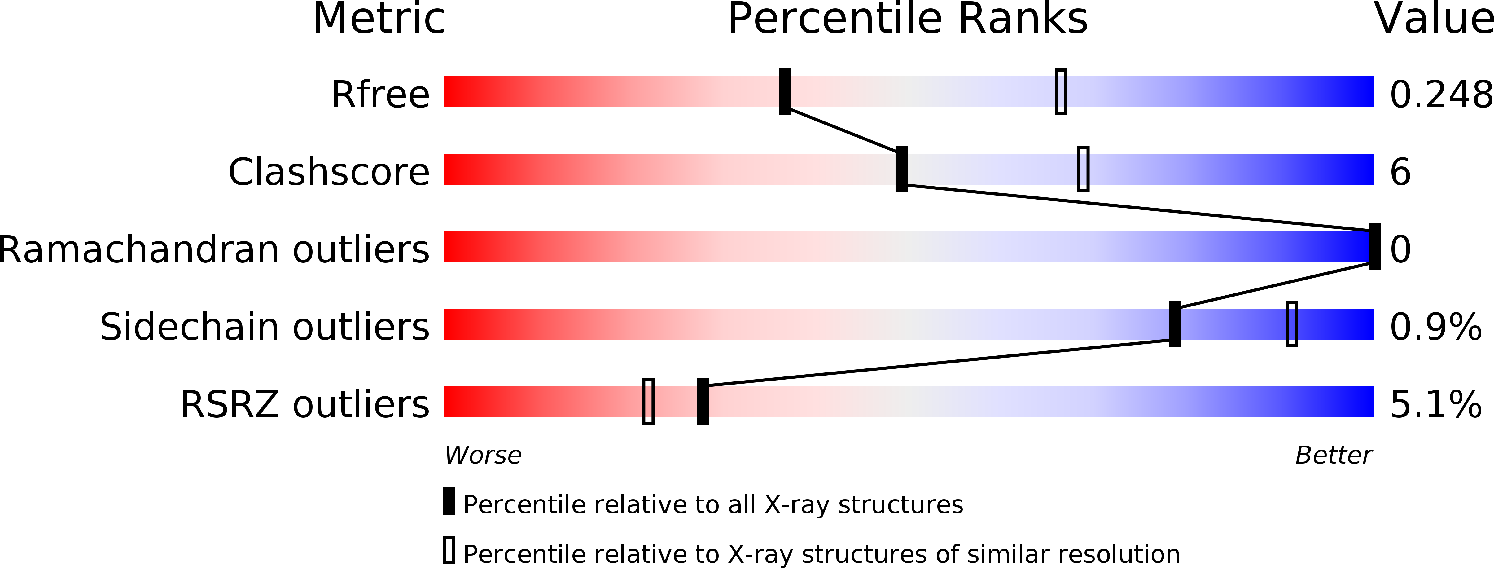
Resolution:
2.60 Å
R-Value Free:
0.24
R-Value Work:
0.19
R-Value Observed:
0.19
Space Group:
P 41 2 2
Macromolecular Entities
Polymer Type:polypeptide(L)
Description:Glutamate receptor ionotropic, kainate 3,Glutamate receptor ionotropic, kainate 3
Chain IDs:A
Chain Length:258
Number of Molecules:1
Biological Source:Rattus norvegicus
Primary Citation
( S)-2-Amino-3-(5-methyl-3-hydroxyisoxazol-4-yl)propanoic Acid (AMPA) and Kainate Receptor Ligands: Further Exploration of Bioisosteric Replacements and Structural and Biological Investigation.
J. Med. Chem. 61 2124 2130 (2018)
PMID: 29451794 DOI: 10.1021/acs.jmedchem.8b00099

Abstact

Starting from 1-4 and 7 structural templates, analogues based on bioisosteric replacements (5a-c vs 1, 2 and 6 vs 7) were synthesized for completing the SAR analysis. Interesting binding properties at GluA2, GluK1, and GluK3 receptors were discovered. The requirements for GluK3 interaction were elucidated by determining the X-ray structures of the GluK3-LBD with 2 and 5c and by computational studies. Antinociceptive potential was demonstrated for GluK1 partial agonist 3 and antagonist 7 (2 mg/kg ip).

Legend

Protein

Chemical

Disease

Primary Citation of related structures