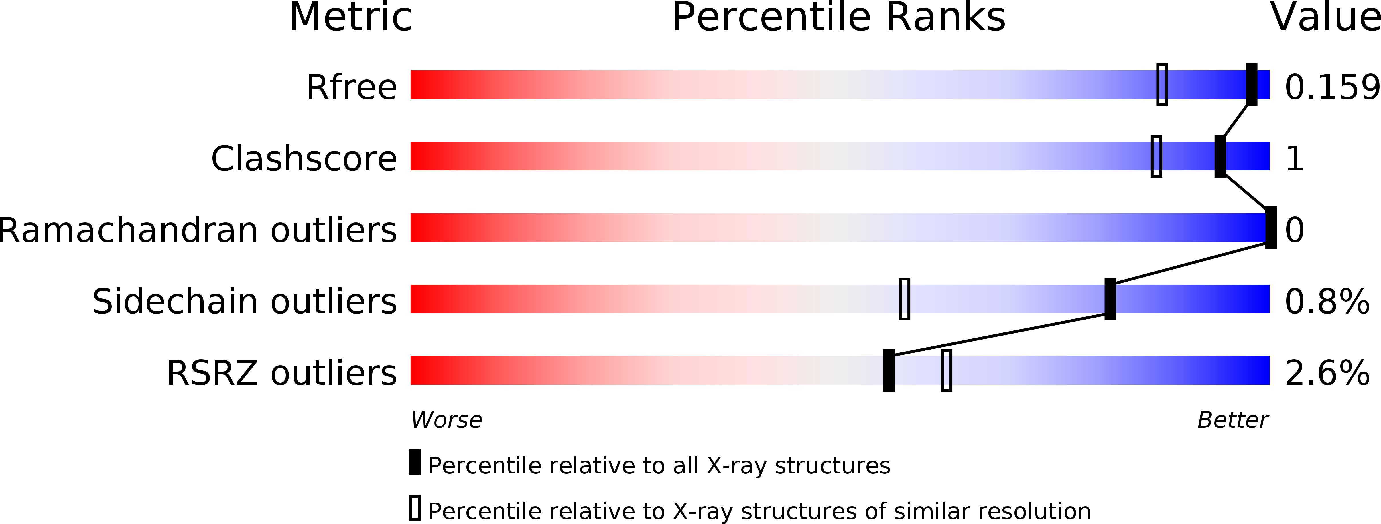
6F0P image
Deposition Date 2017-11-20
Release Date 2018-01-10
Last Version Date 2024-11-06
Entry Detail
PDB ID:
6F0P
Keywords:
Title:
Botulinum neurotoxin A4 Hc domain
Biological Source:
Source Organism:
Host Organism:
Method Details:
Experimental Method:
Resolution:
1.34 Å
R-Value Free:
0.15
R-Value Work:
0.14
R-Value Observed:
0.14
Space Group:
P 21 3
Macromolecular Entities
Structures with similar UniProt ID
Protein Blast
Polymer Type:polypeptide(L)
Molecule:Neurotoxin type A
Chain IDs:A
Chain Length:433
Number of Molecules:1
Biological Source:Clostridium botulinum
Primary Citation
High resolution crystal structures of Clostridium botulinum neurotoxin A3 and A4 binding domains.
J. Struct. Biol. 202 113 117 (2018)
PMID: 29288126 DOI: 10.1016/j.jsb.2017.12.010

Abstact

Clostridium botulinum neurotoxins (BoNTs) cause the life-threatening condition, botulism. However, while they have the potential to cause serious harm, they are increasingly being utilised for therapeutic applications. BoNTs comprise of seven distinct serotypes termed BoNT/A through BoNT/G, with the most widely characterised being sub-serotype BoNT/A1. Each BoNT consists of three structurally distinct domains, a binding domain (HC), a translocation domain (HN), and a proteolytic domain (LC). The HC domain is responsible for the highly specific targeting of the neurotoxin to neuronal cell membranes. Here, we present two high-resolution structures of the binding domain of subtype BoNT/A3 (HC/A3) and BoNT/A4 (HC/A4) at 1.6 Å and 1.34 Å resolution, respectively. The structures of both proteins share a high degree of similarity to other known BoNT HC domains whilst containing some subtle differences, and are of benefit to research into therapeutic neurotoxins with novel characteristics.

Legend

Protein

Chemical

Disease

Primary Citation of related structures