6EYT image
Entry Detail
PDB ID:
6EYT
Keywords:
Title:
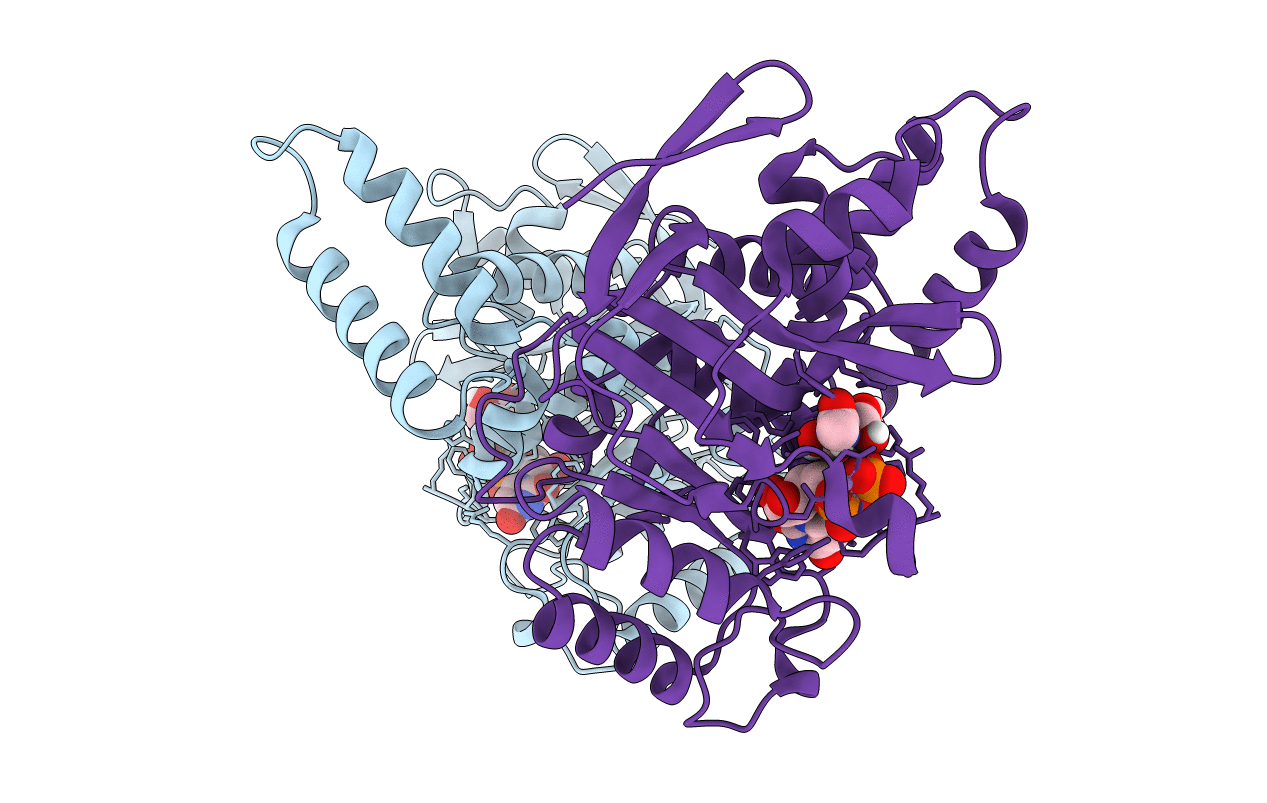
Crystal structure of the Salmonella effector SseK3 in complex with UDP-GlcNAc and Manganese
Biological Source:
Source Organism:
Host Organism:
PDB Version:
Deposition Date:
2017-11-13
Release Date:
2018-02-28
Method Details:
Experimental Method:
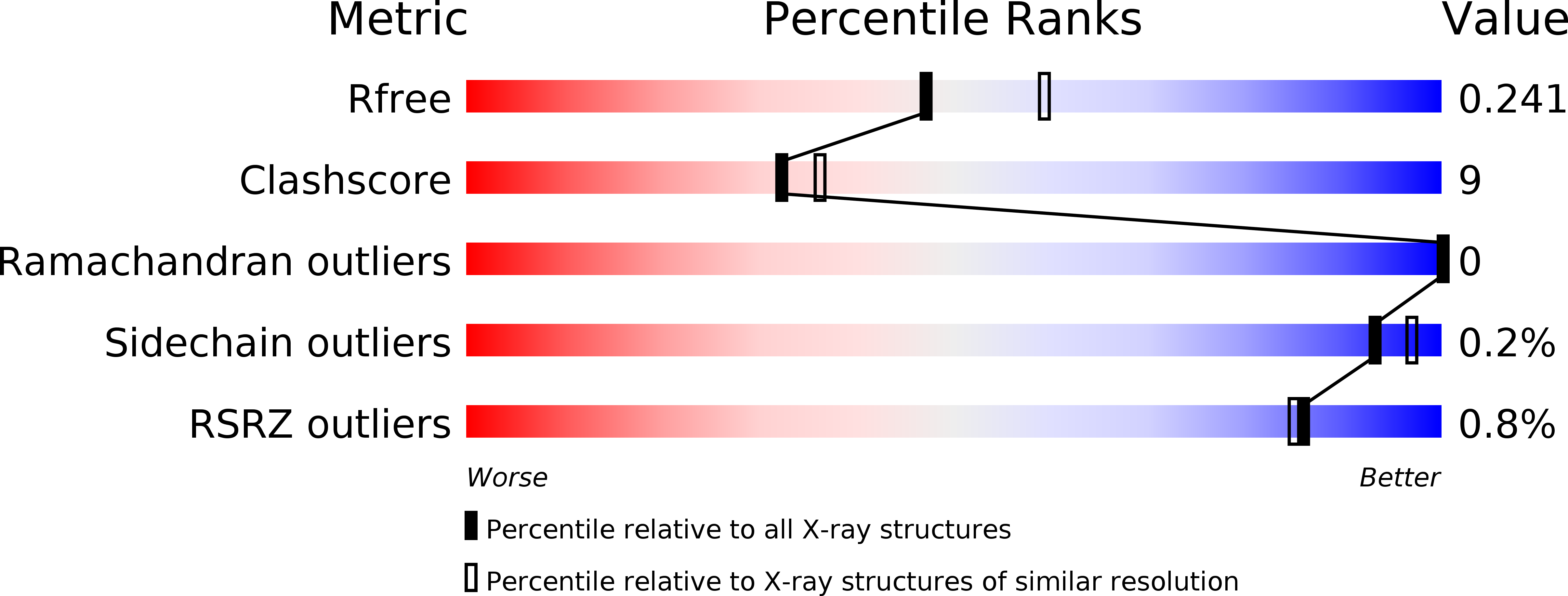
Resolution:
2.21 Å
R-Value Free:
0.24
R-Value Work:
0.19
R-Value Observed:
0.19
Space Group:
P 21 21 21
Macromolecular Entities
Polymer Type:polypeptide(L)
Description:Type III secretion system effector protein
Chain IDs:A, B
Chain Length:327
Number of Molecules:2
Biological Source:Salmonella typhimurium
Primary Citation
Structural basis for the glycosyltransferase activity of theSalmonellaeffector SseK3.
J. Biol. Chem. 293 5064 5078 (2018)
PMID: 29449376 DOI: 10.1074/jbc.RA118.001796

Abstact

The Salmonella-secreted effector SseK3 translocates into host cells, targeting innate immune responses, including NF-kappaB activation. SseK3 is a glycosyltransferase that transfers an N-acetylglucosamine (GlcNAc) moiety onto the guanidino group of a target arginine, modulating host cell function. However, a lack of structural information has precluded elucidation of the molecular mechanisms in arginine and GlcNAc selection. We report here the crystal structure of SseK3 in its apo form and in complex with hydrolyzed UDP-GlcNAc. SseK3 possesses the typical glycosyltransferase type-A (GT-A)-family fold and the metal-coordinating DXD motif essential for ligand binding and enzymatic activity. Several conserved residues were essential for arginine GlcNAcylation and SseK3-mediated inhibition of NF-kappaB activation. Isothermal titration calorimetry revealed SseK3's preference for manganese coordination. The pattern of interactions in the substrate-bound SseK3 structure explained the selection of the primary ligand. Structural rearrangement of the C-terminal residues upon ligand binding was crucial for SseK3's catalytic activity, and NMR analysis indicated that SseK3 has limited UDP-GlcNAc hydrolysis activity. The release of free N-acetyl alpha-d-glucosamine, and the presence of the same molecule in the SseK3 active site, classified it as a retaining glycosyltransferase. A glutamate residue in the active site suggested a double-inversion mechanism for the arginine N-glycosylation reaction. Homology models of SseK1, SseK2, and the Escherichia coli orthologue NleB1 reveal differences in the surface electrostatic charge distribution, possibly accounting for their diverse activities. This first structure of a retaining GT-A arginine N-glycosyltransferase provides an important step toward a better understanding of this enzyme class and their roles as bacterial effectors.

Legend

Protein

Chemical

Disease

Primary Citation of related structures