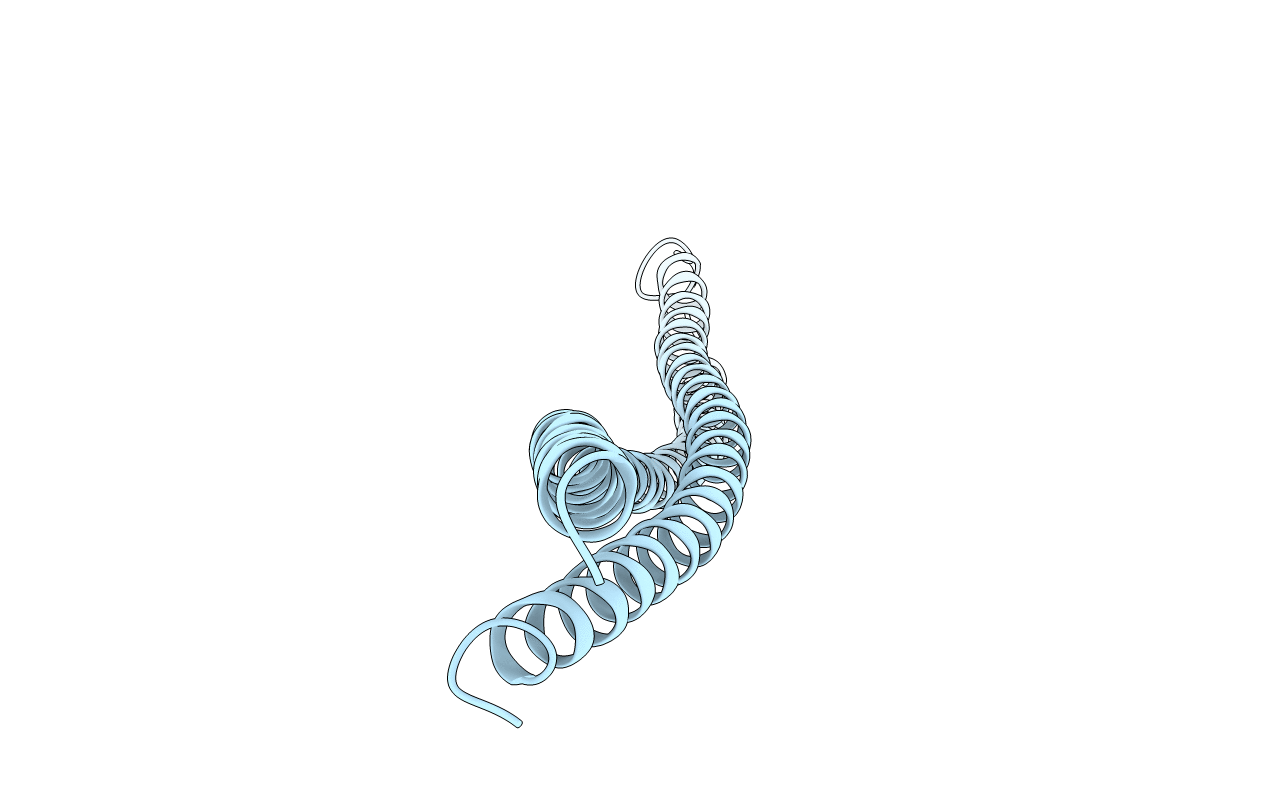
6EWY image
Entry Detail
PDB ID:
6EWY
Title:
RipA Peptidoglycan hydrolase (Rv1477, Mycobacterium tuberculosis) N-terminal domain
Biological Source:
PDB Version:
Deposition Date:
2017-11-07
Release Date:
2018-05-02
Method Details:
Experimental Method:
Resolution:
2.20 Å
R-Value Free:
0.26
R-Value Work:
0.22
R-Value Observed:
0.23
Space Group:
C 2 2 2
Macromolecular Entities
Polymer Type:polypeptide(L)
Description:Peptidoglycan endopeptidase RipA
Chain IDs:A
Chain Length:219
Number of Molecules:1
Biological Source:Mycobacterium tuberculosis (strain ATCC 25618 / H37Rv)
Primary Citation
The structure of the N-terminal module of the cell wall hydrolase RipA and its role in regulating catalytic activity.
Proteins 86 912 923 (2018)
PMID: 29722065 DOI: 10.1002/prot.25523

Abstact

RipA plays a vital role during cell division of Mycobacterium tuberculosis by degrading the cell wall peptidoglycan at the septum, allowing daughter cell separation. The peptidoglycan degrading activity relies on the NlpC/P60 domain, and as it is potentially harmful when deregulated, spatial and temporal control is necessary in this process. The N-terminal domain of RipA has been proposed to play an inhibitory role blocking the C-terminal NlpC/P60 domain. Accessibility of the active site cysteine residue is however not limited by the presence of the N-terminal domain, but by the lid-module of the inter-domain linker, which is situated in the peptide binding groove of the crystal structures of the catalytic domain. The 2.2 Å resolution structure of the N-terminal domain, determined by Se-SAD phasing, reveals an all-α-fold with 2 long α-helices, and shows similarity to bacterial periplasmic protein domains with scaffold-building role. Size exclusion chromatography and SAXS experiments are consistent with dimer formation of this domain in solution. The SAXS data from the periplasmic two-domain RipA construct suggest a rigid baton-like structure of the N-terminal module, with the catalytic domain connected by a 24 residue long flexible linker. This flexible linker allows for a catalytic zone, which is part of the spatiotemporal control of peptidoglycan degradation.

Legend

Protein

Chemical

Disease

Primary Citation of related structures