
Deposition Date
2017-11-01
Release Date
2017-12-13
Last Version Date
2024-01-17
Entry Detail
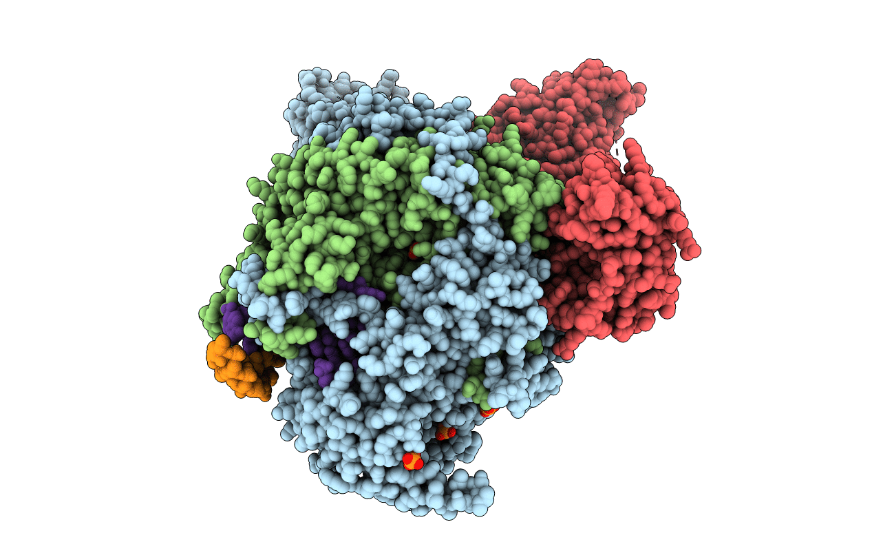
PDB ID:
6EVK
Keywords:
Title:
Crystal structure of bat influenza A/H17N10 polymerase with viral RNA promoter and cap analogue m7GTP
Biological Source:
Source Organism(s):
Expression System(s):
Method Details:
Experimental Method:
Resolution:
2.90 Å
R-Value Free:
0.27
R-Value Work:
0.23
R-Value Observed:
0.23
Space Group:
C 1 2 1
