Deposition Date
2017-10-27
Release Date
2018-02-21
Last Version Date
2024-10-16
Method Details:
Experimental Method:
Resolution:
1.08 Å
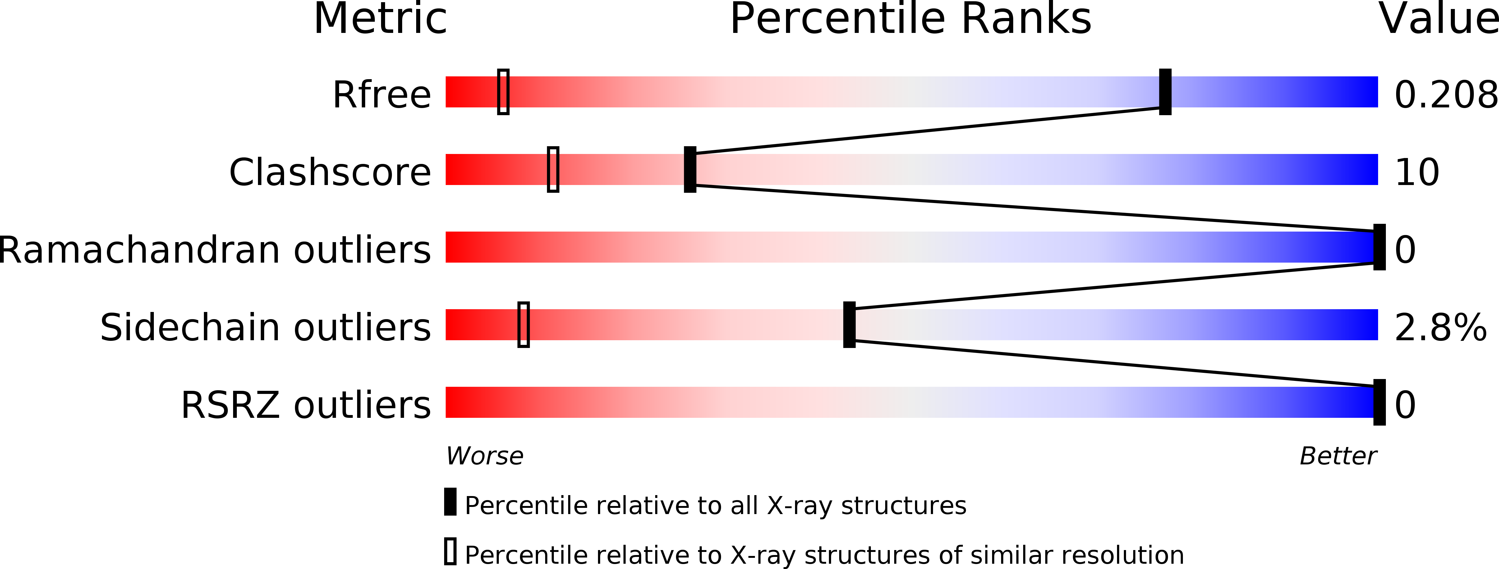
R-Value Free:
0.21
R-Value Work:
0.14
R-Value Observed:
0.15
Space Group:
P 1 21 1