6ET6 image
Deposition Date 2017-10-25
Release Date 2018-09-05
Last Version Date 2024-01-17
Entry Detail
PDB ID:
6ET6
Title:
Crystal structure of muramidase from Acinetobacter baumannii AB 5075UW prophage
Biological Source:
Source Organism:
Host Organism:
Method Details:
Experimental Method:
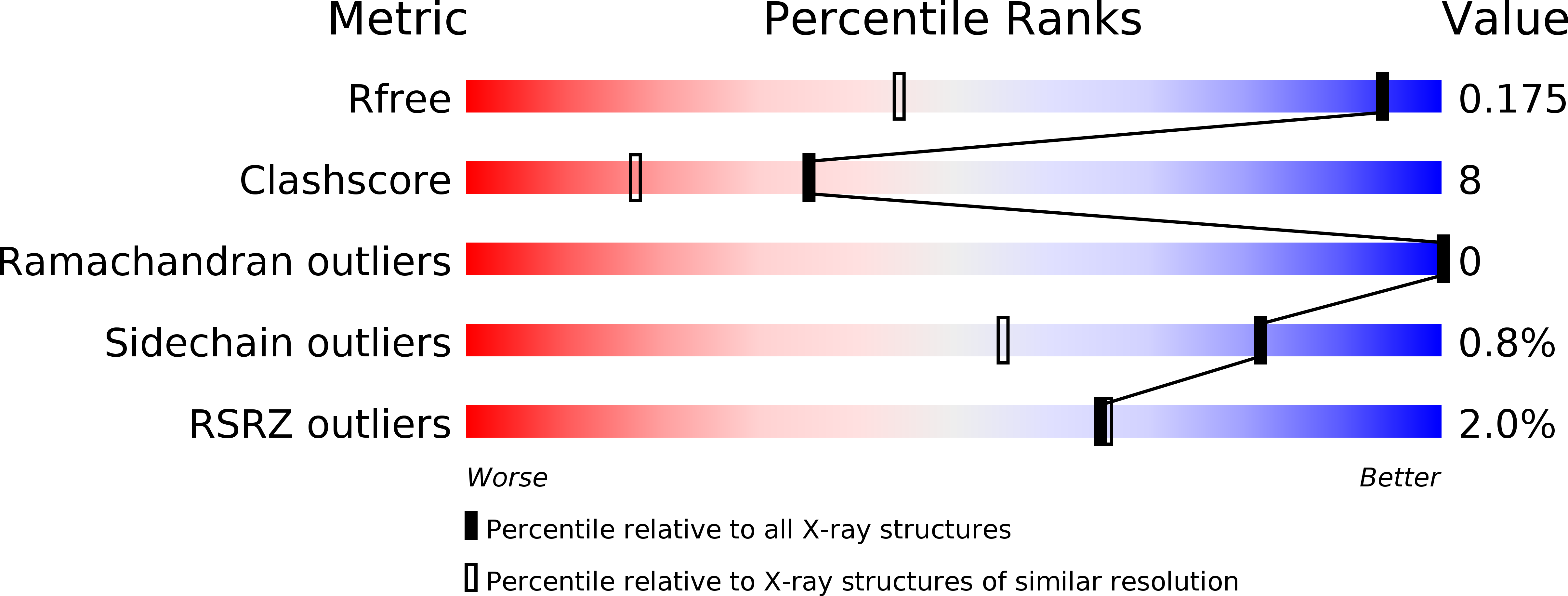
Resolution:
1.20 Å
R-Value Free:
0.16
R-Value Work:
0.14
R-Value Observed:
0.14
Space Group:
P 2 21 21
Macromolecular Entities
Structures with similar UniProt ID
Protein Blast
Polymer Type:polypeptide(L)
Molecule:Lysozyme
Gene (Uniprot):ABUW_1326, EA686_25815
Chain IDs:A
Chain Length:196
Number of Molecules:1
Biological Source:Acinetobacter baumannii
Primary Citation
Structure of anAcinetobacterBroad-Range Prophage Endolysin Reveals a C-Terminal alpha-Helix with the Proposed Role in Activity against Live Bacterial Cells.
Viruses 10 ? ? (2018)
PMID: 29882827 DOI: 10.3390/v10060309
Primary Citation of related structures