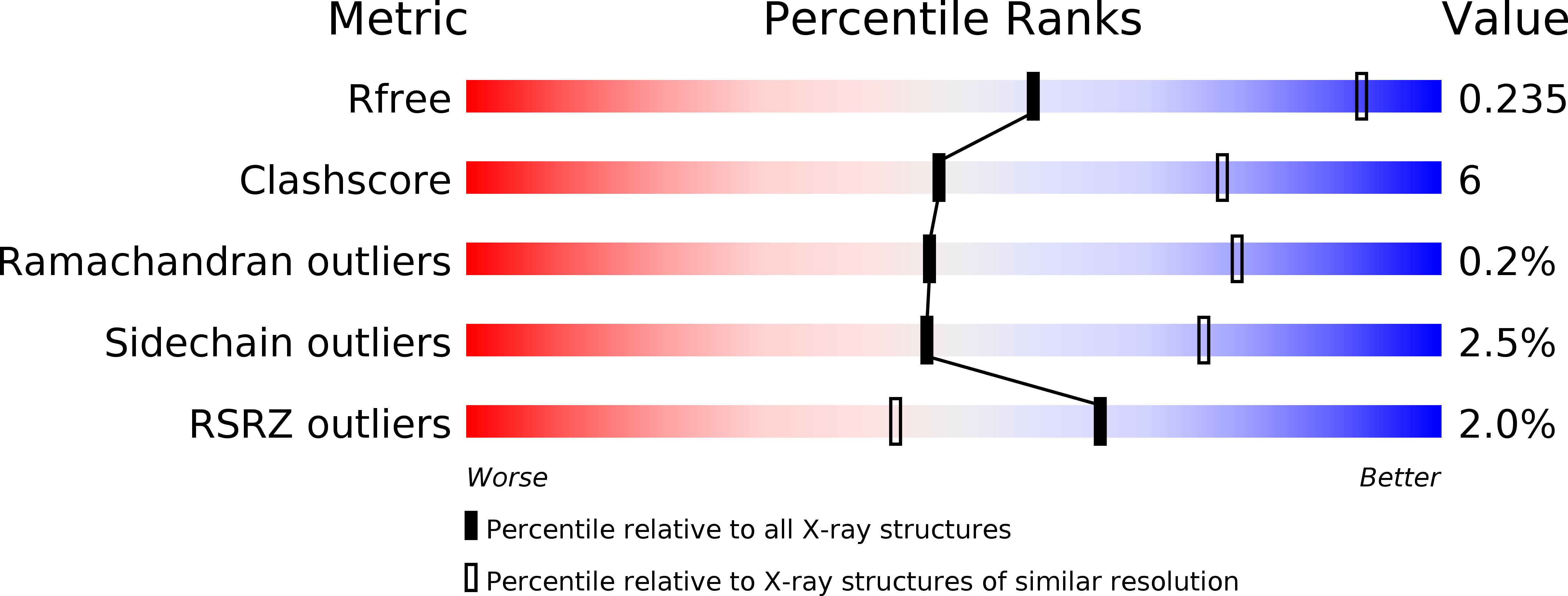
6EQI image
Deposition Date 2017-10-13
Release Date 2017-11-08
Last Version Date 2024-10-23
Entry Detail
PDB ID:
6EQI
Keywords:
Title:
Structure of PINK1 bound to ubiquitin
Biological Source:
Source Organism:
Host Organism:
Method Details:
Experimental Method:
Resolution:
3.10 Å
R-Value Free:
0.23
R-Value Work:
0.20
R-Value Observed:
0.20
Space Group:
P 31 2 1
Macromolecular Entities
Structures with similar UniProt ID
Protein Blast
Polymer Type:polypeptide(L)
Molecule:Ubiquitin
Gene (Uniprot):UBC
Mutagens:T66V L67N
Chain IDs:A
Chain Length:76
Number of Molecules:1
Biological Source:Homo sapiens
Polymer Type:polypeptide(L)
Molecule:Nb696
Chain IDs:B
Chain Length:134
Number of Molecules:1
Biological Source:Lama glama
Structures with similar UniProt ID
Protein Blast
Polymer Type:polypeptide(L)
Molecule:Serine/threonine-protein kinase PINK1, putative
Gene (Uniprot):Pink1
Chain IDs:C
Chain Length:433
Number of Molecules:1
Biological Source:Pediculus humanus corporis
Modified Residue
Compound ID Chain ID Parent Comp ID Details 2D Image
SEP C SER modified residue
TPO C THR modified residue
Ligand Molecules
Primary Citation
Structure of PINK1 in complex with its substrate ubiquitin.
Nature 552 51 56 (2017)
PMID: 29160309 DOI: 10.1038/nature24645

Abstact

Autosomal-recessive juvenile Parkinsonism (AR-JP) is caused by mutations in a number of PARK genes, in particular the genes encoding the E3 ubiquitin ligase Parkin (PARK2, also known as PRKN) and its upstream protein kinase PINK1 (also known as PARK6). PINK1 phosphorylates both ubiquitin and the ubiquitin-like domain of Parkin on structurally protected Ser65 residues, triggering mitophagy. Here we report a crystal structure of a nanobody-stabilized complex containing Pediculus humanus corporis (Ph)PINK1 bound to ubiquitin in the 'C-terminally retracted' (Ub-CR) conformation. The structure reveals many peculiarities of PINK1, including the architecture of the C-terminal region, and reveals how the N lobe of PINK1 binds ubiquitin via a unique insertion. The flexible Ser65 loop in the Ub-CR conformation contacts the activation segment, facilitating placement of Ser65 in a phosphate-accepting position. The structure also explains how autophosphorylation in the N lobe stabilizes structurally and functionally important insertions, and reveals the molecular basis of AR-JP-causing mutations, some of which disrupt ubiquitin binding.

Legend

Protein

Chemical

Disease

Primary Citation of related structures