6EO9 image
Deposition Date 2017-10-09
Release Date 2017-12-13
Last Version Date 2024-11-20
Entry Detail
PDB ID:
6EO9
Keywords:
Title:
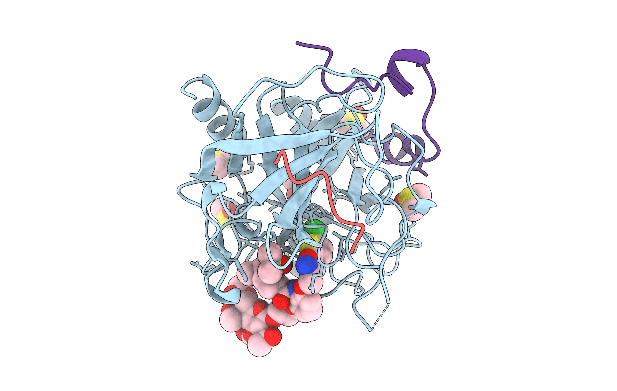
Crystal structure of thrombin in complex with a novel glucose-conjugated potent inhibitor
Biological Source:
Source Organism:
Homo sapiens (Taxon ID: 9606)
Hirudo medicinalis (Taxon ID: 6421)
Host Organism:
Method Details:
Experimental Method:
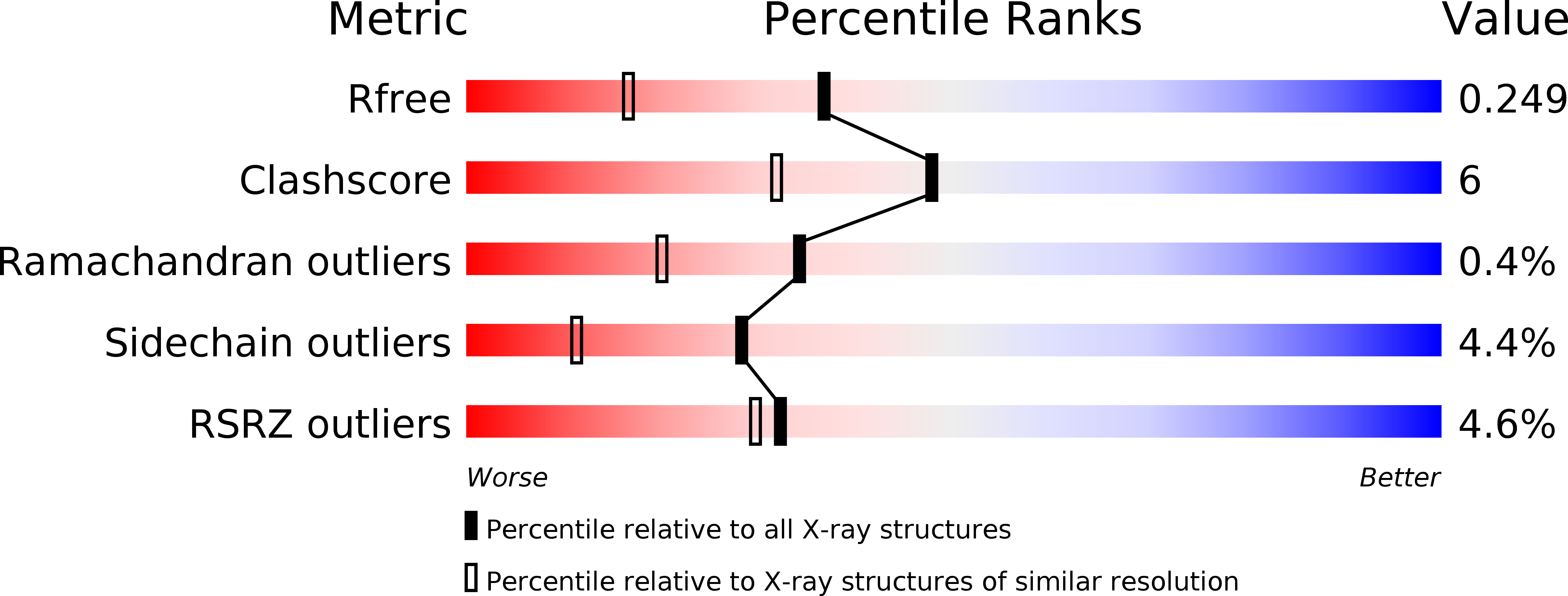
Resolution:
1.84 Å
R-Value Free:
0.24
R-Value Work:
0.19
R-Value Observed:
0.20
Space Group:
C 1 2 1
Macromolecular Entities
Polymer Type:polypeptide(L)
Molecule:Prothrombin
Gene (Uniprot):F2
Chain IDs:A (auth: H)
Chain Length:259
Number of Molecules:1
Biological Source:Homo sapiens
Polymer Type:polypeptide(L)
Molecule:Hirudin variant-2
Chain IDs:C (auth: I)
Chain Length:12
Number of Molecules:1
Biological Source:Hirudo medicinalis
Polymer Type:polypeptide(L)
Molecule:Prothrombin
Gene (Uniprot):F2
Chain IDs:B (auth: L)
Chain Length:36
Number of Molecules:1
Biological Source:Homo sapiens
Modified Residue
Compound ID Chain ID Parent Comp ID Details 2D Image
TYS C TYR modified residue
Primary Citation
How a beta-D-glucoside side chain enhances binding affinity to thrombin of inhibitors bearing 2-chlorothiophene as P1 moiety: crystallography, fragment deconstruction study, and evaluation of antithrombotic properties.
J. Med. Chem. 57 8563 8575 (2014)
PMID: 25268757 DOI: 10.1021/jm5010754

Abstact

The β-d-glucose-containing compound 3, bearing 2-chlorothiophene and 1-isopropylpiperidine moieties as binders of the S1 and S4 pockets, respectively, proved to be potent competitive inhibitor of factor Xa (fXa, Ki = 0.090 nM) and thrombin (fIIa, Ki = 100 nM). The potency of 3 increases, over the parent compound 1, against fIIa (110-fold), much more than against fXa (7-fold). Experimental deconstruction of 3 into smaller fragments revealed a binding cooperativity of the P3/P4 and propylene-linked β-d-glucose fragments, stronger in fIIa (15.5 kJ·mol(-1)) than in fXa (2.8 kJ·mol(-1)). The crystal structure of human fIIa in complex with 3 revealed a binding mode including a strong H-bond network between the glucose O1', O3', and O5' and two critical residues, namely R221a and K224, belonging to the Na(+)-binding site which may allosterically perturb the specificity sites. The potential of 3 as antithrombotic agent was supported by its ability to inhibit thrombin generation and to stimulate fibrinolysis at submicromolar concentration.

Legend

Protein

Chemical

Disease

Primary Citation of related structures