Deposition Date
2017-10-09
Release Date
2017-10-25
Last Version Date
2025-10-01
Entry Detail
PDB ID:
6EO6
Keywords:
Title:
X-ray structure of the complex between human alpha-thrombin and modified 15-mer DNA aptamer containing 5-(3-(2-(1H-indol-3-yl)acetamide-N-yl)-1-propen-1-yl)-2'-deoxyuridine residue
Biological Source:
Source Organism(s):
synthetic construct (Taxon ID: 32630)
Homo sapiens (Taxon ID: 9606)
Homo sapiens (Taxon ID: 9606)
Method Details:
Experimental Method:
Resolution:
1.69 Å
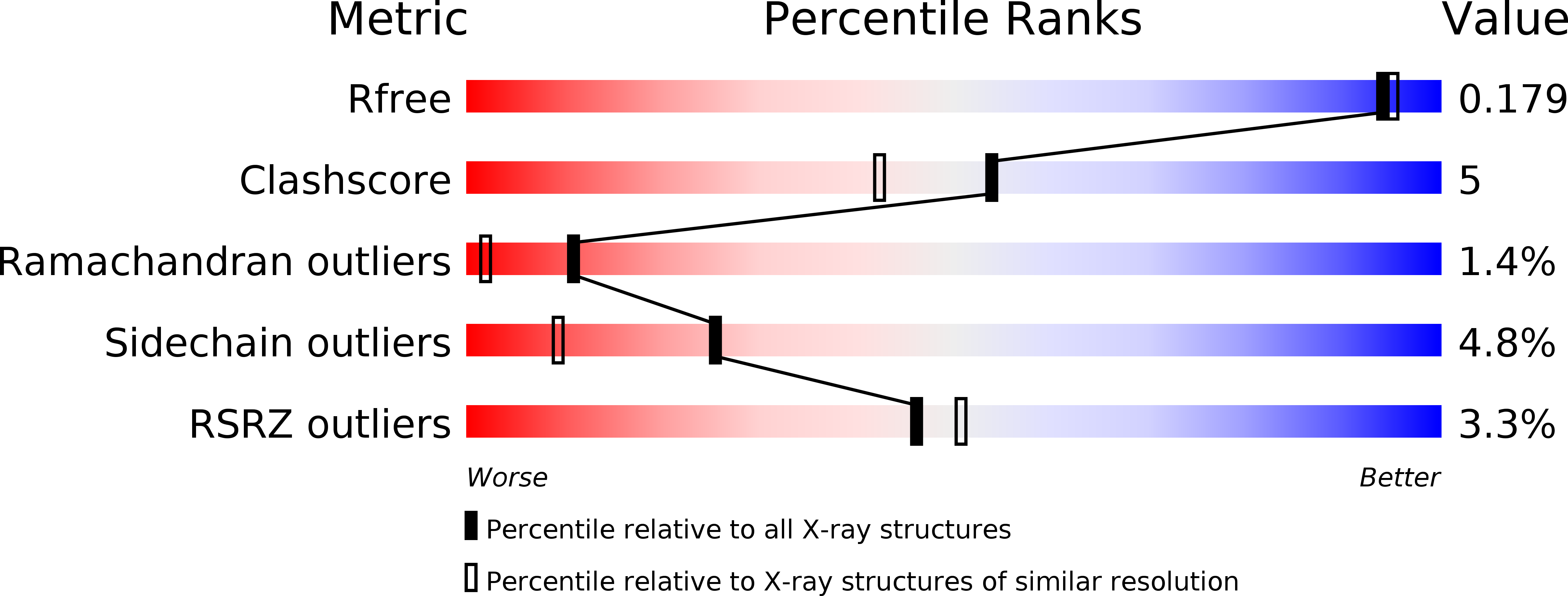
R-Value Free:
0.16
R-Value Work:
0.14
R-Value Observed:
0.14
Space Group:
P 32 2 1