
Deposition Date
2017-09-29
Release Date
2018-06-20
Last Version Date
2024-01-17
Entry Detail
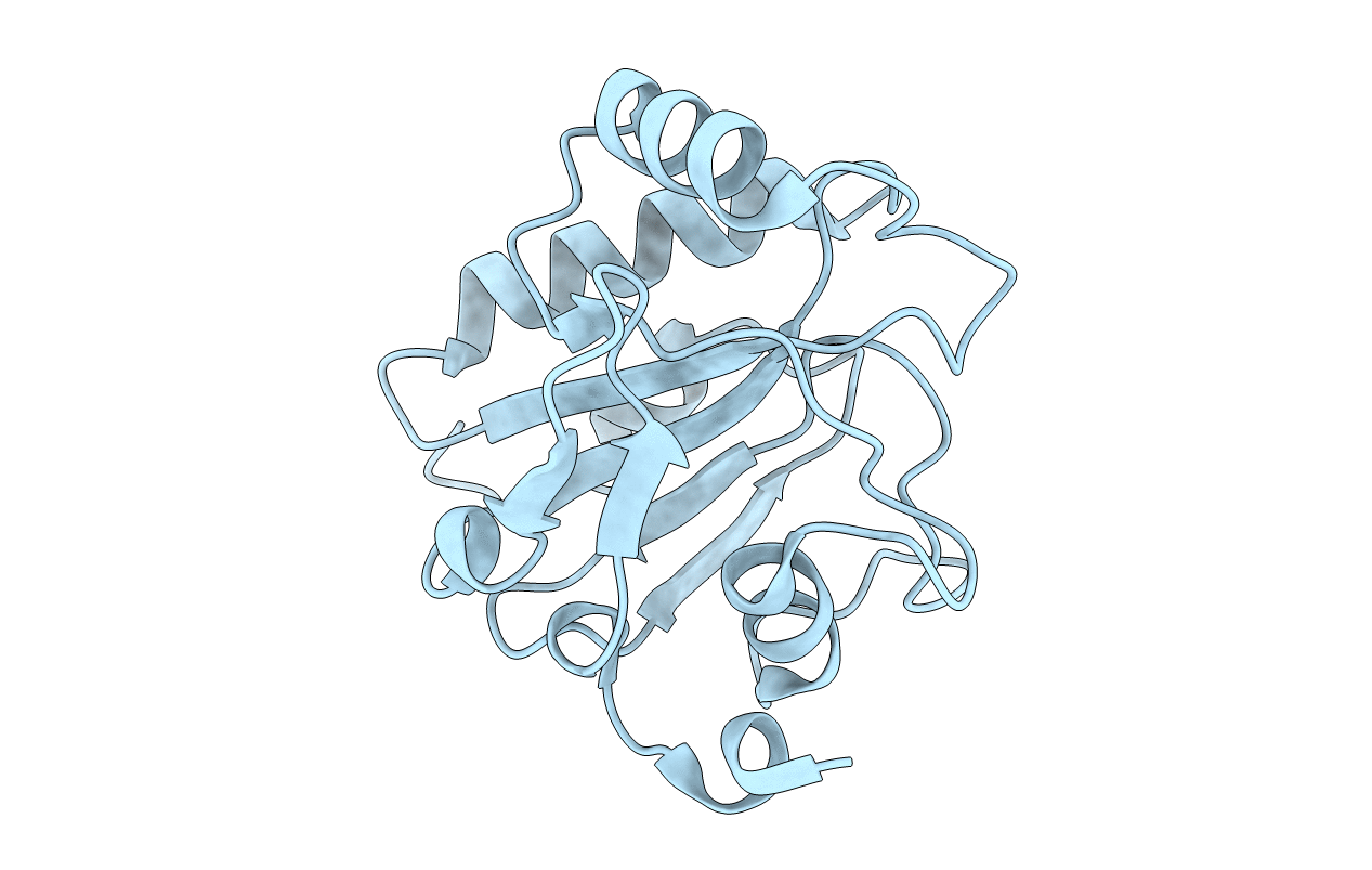
PDB ID:
6ELW
Keywords:
Title:
High resolution structure of selenocysteine containing human GPX4
Biological Source:
Source Organism(s):
Homo sapiens (Taxon ID: 9606)
Expression System(s):
Method Details:
Experimental Method:
Resolution:
1.30 Å
R-Value Free:
0.15
R-Value Work:
0.12
R-Value Observed:
0.12
Space Group:
P 1 21 1
