6EHO image
Deposition Date 2017-09-14
Release Date 2017-12-06
Last Version Date 2024-11-06
Entry Detail
PDB ID:
6EHO
Keywords:
Title:
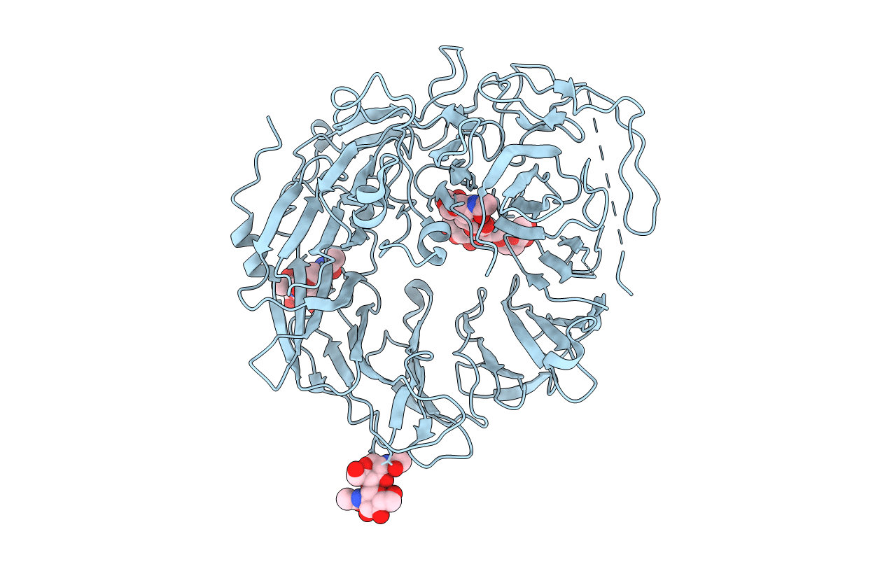
Dimer of the Sortilin Vps10p domain at low pH
Biological Source:
Source Organism:
Homo sapiens (Taxon ID: 9606)
Host Organism:
Method Details:
Experimental Method:
Resolution:
3.50 Å
R-Value Free:
0.27
R-Value Work:
0.23
R-Value Observed:
0.23
Space Group:
I 21 3
Macromolecular Entities
Polymer Type:polypeptide(L)
Molecule:Sortilin
Gene (Uniprot):SORT1
Mutations:R43G , R44G
Chain IDs:A
Chain Length:723
Number of Molecules:1
Biological Source:Homo sapiens
Ligand Molecules
Primary Citation
Acidic Environment Induces Dimerization and Ligand Binding Site Collapse in the Vps10p Domain of Sortilin.
Structure 25 1809 1819.e3 (2017)
PMID: 29107483 DOI: 10.1016/j.str.2017.09.015

Abstact

Sortilin is a neuronal receptor involved in transmembrane signaling, endocytosis, and intracellular sorting of proteins. It cycles through a number of cellular compartments where it encounters various acidic conditions. The crystal structure of the sortilin ectodomain has previously been determined at neutral pH. Here, we present the 3.5-Å resolution crystal structure of sortilin at pH 5.5, which represents an environment similar to that of late endosomes, where ligands are released. The structure reveals an overall distortion of the 10-bladed β-propeller domain. This distortion and specific conformational changes, caused by protonation of a number of histidine residues, render the currently known binding sites unavailable for ligand binding. Access to the binding sites is furthermore blocked by a reversible and pH-dependent formation of tight sortilin dimers, also confirmed by electron microscopy, size-exclusion chromatography, and mutational studies. This study reveals how sortilin binding sites are disrupted and explains pH-dependent ligand affinity.

Legend

Protein

Chemical

Disease

Primary Citation of related structures