6EGY image
Deposition Date 2017-09-12
Release Date 2018-10-10
Last Version Date 2024-10-16
Entry Detail
PDB ID:
6EGY
Title:
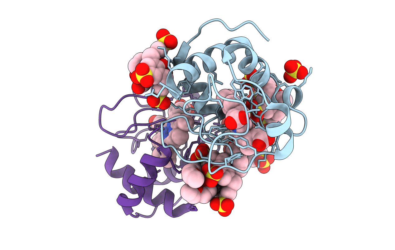
Crystal structure of cytochrome c in complex with mono-PEGylated sulfonatocalix[4]arene
Biological Source:
Source Organism:
Method Details:
Experimental Method:
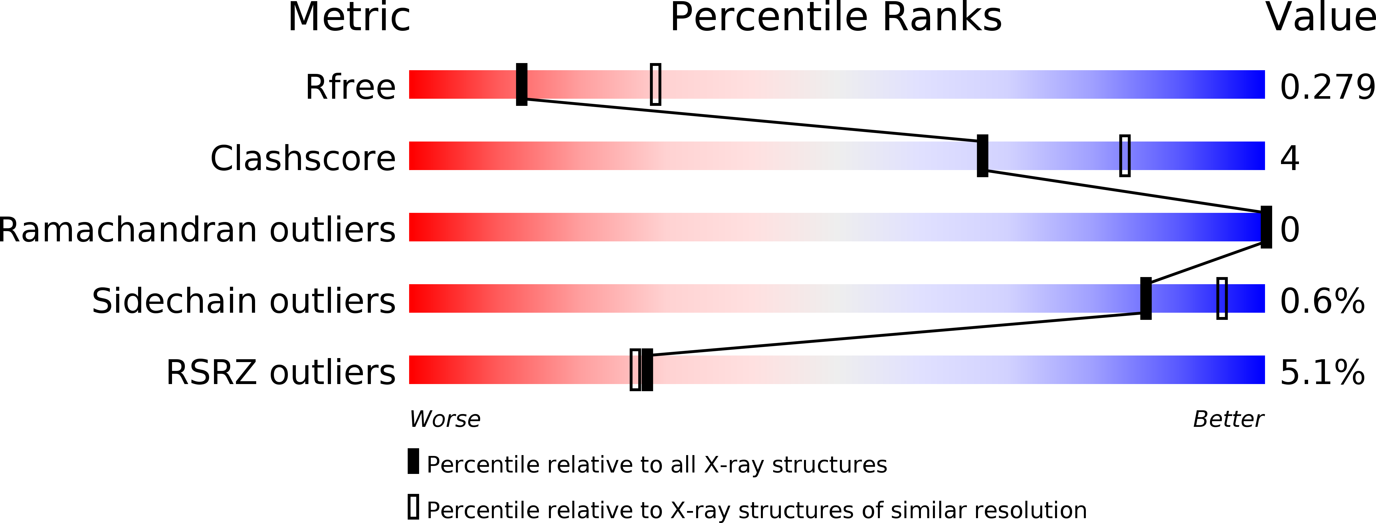
Resolution:
2.70 Å
R-Value Free:
0.27
R-Value Work:
0.22
R-Value Observed:
0.22
Space Group:
I 41 3 2
Macromolecular Entities
Structures with similar UniProt ID
Protein Blast
Polymer Type:polypeptide(L)
Molecule:Cytochrome c iso-1
Gene (Uniprot):CYC1
Mutagens:C102T
Chain IDs:A, B
Chain Length:108
Number of Molecules:2
Biological Source:Saccharomyces cerevisiae
Primary Citation
Noncovalent PEGylation via Sulfonatocalix[4]arene-A Crystallographic Proof.
Bioconjug.Chem. 29 3999 4003 (2018)
PMID: 30445810 DOI: 10.1021/acs.bioconjchem.8b00769

Abstact

Noncovalent or supramolecular PEGylation, in combination with the site of administration, has great potential to increase the half-life of therapeutic proteins. To date, a variety of noncovalent PEGylation strategies have been devised. However, questions remain concerning the nature of the protein-PEG interaction. Here, we report structural analyses of a model system that comprised the lysine-rich cytochrome c and two PEGylated variants of sulfonatocalix[4]arene. Complex formation was characterized in solution by NMR spectroscopy. It was found that mono- or di-PEGylated sulfonatocalix[4]arene bound the protein similar to the parent calixarene. X-ray crystal structures at <2.7 Å resolution of the PEGylated derivatives in complex with cytochrome c revealed that the PEG chains were mostly disordered or encapsulated within the calixarene cavity. These results suggest that there was minimal interaction between the PEG and the protein surface, providing further evidence in favor of PEG maintaining a random coil conformation.

Legend

Protein

Chemical

Disease

Primary Citation of related structures