6EEV image
Entry Detail
PDB ID:
6EEV
Keywords:
Title:
Structure of class II HMG-CoA reductase from Delftia acidovorans with mevalonate bound
Biological Source:
Source Organism:
Host Organism:
PDB Version:
Deposition Date:
2018-08-15
Release Date:
2018-09-26
Method Details:
Experimental Method:
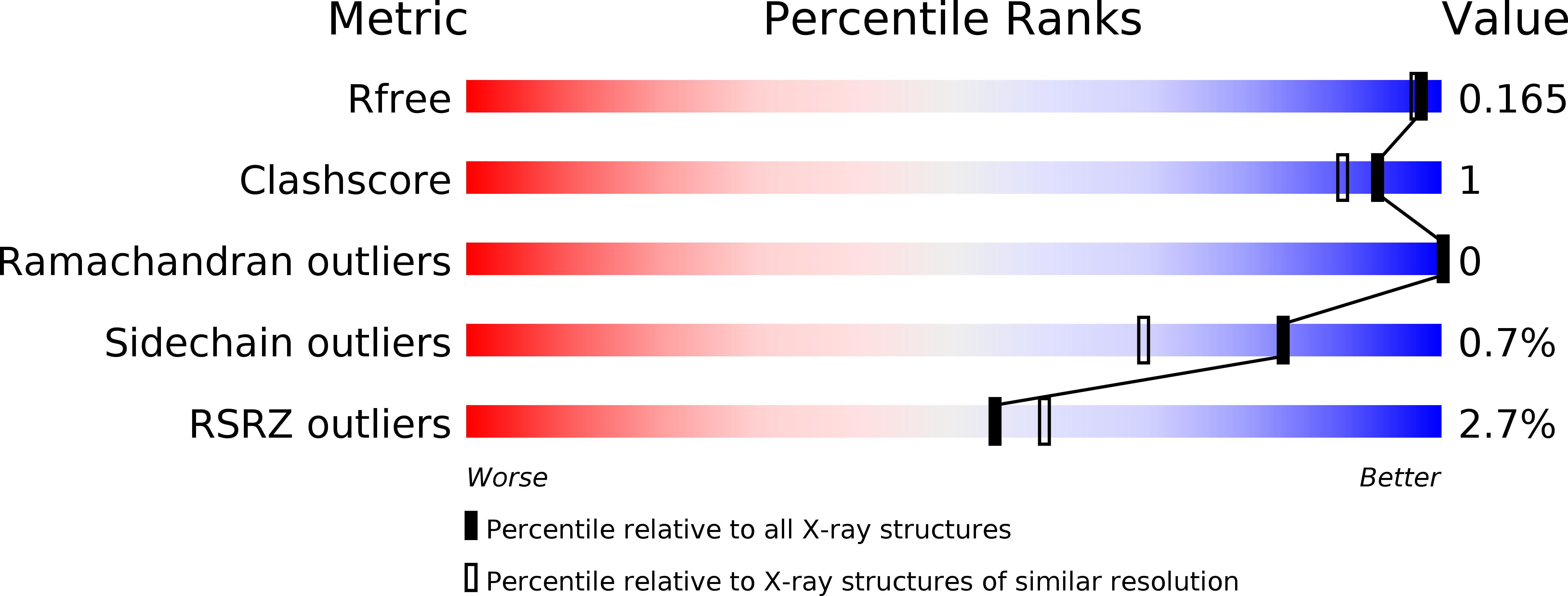
Resolution:
1.49 Å
R-Value Free:
0.16
R-Value Work:
0.13
R-Value Observed:
0.13
Space Group:
P 3 2 1
Macromolecular Entities
Polymer Type:polypeptide(L)
Description:3-hydroxy-3-methylglutaryl coenzyme A reductase
Chain IDs:A
Chain Length:429
Number of Molecules:1
Biological Source:Delftia acidovorans
Peptide-like Molecules
PRD_900003
Primary Citation
New Crystallographic Snapshots of Large Domain Movements in Bacterial 3-Hydroxy-3-methylglutaryl Coenzyme A Reductase.
Biochemistry 57 5715 5725 (2018)
PMID: 30199631 DOI: 10.1021/acs.biochem.8b00869

Abstact

The enzyme 3-hydroxy-3-methylglutaryl coenzyme A (HMG-CoA) reductase (HMGR) catalyzes the first committed step of the mevalonate pathway, which is used across biology in the biosynthesis of countless metabolites. HMGR consumes 2 equiv of the cofactor NAD(P)H to perform the four-electron reduction of HMG-CoA to mevalonate toward the production of steroids and isoprenoids, the largest class of natural products. Recent structural data have shown that HMGR contains a highly mobile C-terminal domain (CTD) that is believed to adopt many different conformations to permit binding and dissociation of the substrate, cofactors, and products at specific points during the reaction cycle. Here, we have characterized the HMGR from Delftia acidovorans as an NADH-specific enzyme and determined crystal structures of the enzyme in unbound, mevalonate-bound, and NADH- and citrate-bound states. Together, these structures depict ligand binding in both the active site and the cofactor-binding site while illustrating how a conserved helical motif confers NAD(P)H cofactor specificity. Unexpectedly, the NADH-bound structure also reveals a new conformation of the CTD, in which the domain has "flipped" upside-down, while directly binding the cofactor. By capturing these structural snapshots, this work not only expands the known range of HMGR domain movement but also provides valuable insight into the catalytic mechanism of this biologically important enzyme.

Legend

Protein

Chemical

Disease

Primary Citation of related structures