6EEL image
Deposition Date 2018-08-14
Release Date 2019-05-22
Last Version Date 2023-10-11
Entry Detail
PDB ID:
6EEL
Title:
Crystal Structure of Myoferlin C2A with divalent cation
Biological Source:
Source Organism:
Homo sapiens (Taxon ID: 9606)
Host Organism:
Method Details:
Experimental Method:
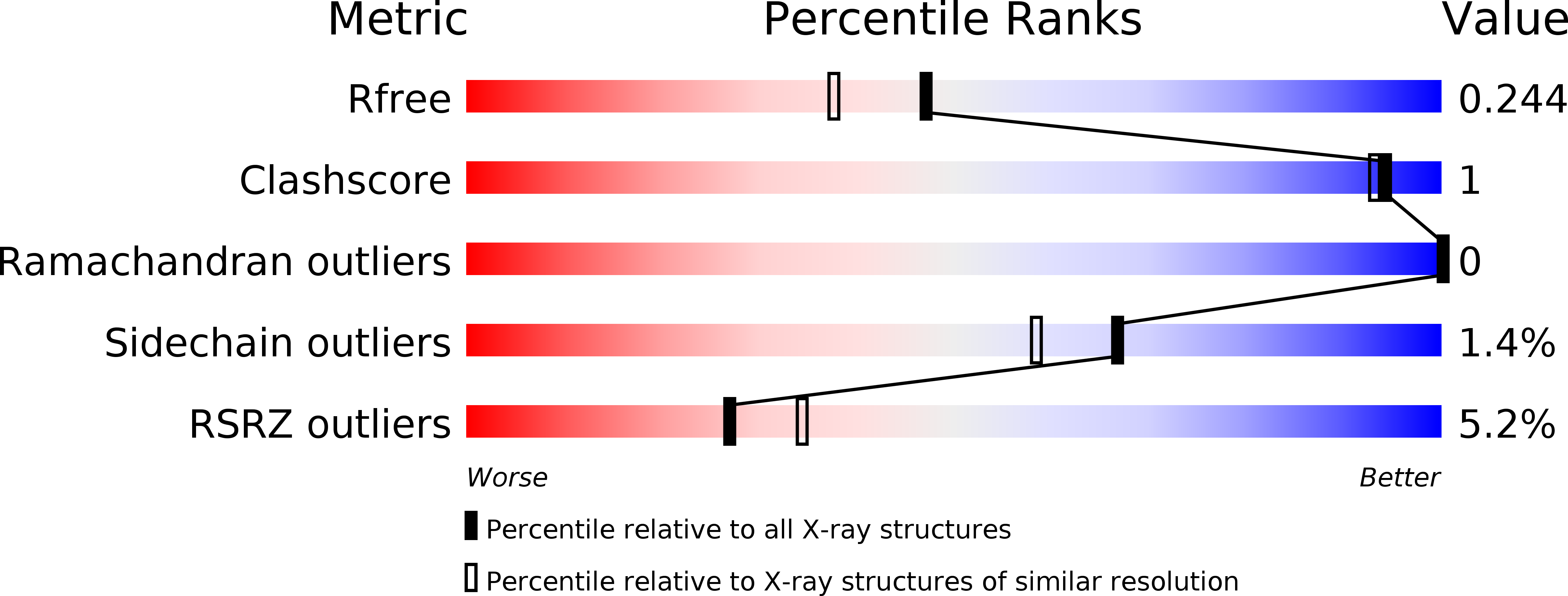
Resolution:
1.93 Å
R-Value Free:
0.23
R-Value Work:
0.18
R-Value Observed:
0.18
Space Group:
P 21 21 21
Macromolecular Entities
Polymer Type:polypeptide(L)
Molecule:Myoferlin
Gene (Uniprot):MYOF
Chain IDs:A, B, C
Chain Length:129
Number of Molecules:3
Biological Source:Homo sapiens
Ligand Molecules
Primary Citation
Structural Basis for the Distinct Membrane Binding Activity of the Homologous C2A Domains of Myoferlin and Dysferlin.
J.Mol.Biol. 431 2112 2126 (2019)
PMID: 31004665 DOI: 10.1016/j.jmb.2019.04.006

Abstact

Dysferlin has been implicated in acute membrane repair processes, whereas myoferlin's activity is maximal during the myoblast fusion stage of early skeletal muscle cell development. Both proteins are similar in size and domain structure; however, despite the overall similarity, myoferlin's known physiological functions do not overlap with those of dysferlin. Here we present for the first time the X-ray crystal structure of human myoferlin C2A to 1.9 Å resolution bound to two divalent cations, and compare its three-dimensional structure and membrane binding activities to that of dysferlin C2A. We find that while dysferlin C2A binds membranes in a Ca2+-dependent manner, Ca2+ binding was the rate-limiting kinetic step for this interaction. Myoferlin C2A, on the other hand, binds two calcium ions with an affinity 3-fold lower than that of dysferlin C2A; and, surprisingly, myoferlin C2A binds only marginally to phospholipid mixtures with a high fraction of phosphatidylserine.

Legend

Protein

Chemical

Disease

Primary Citation of related structures