Deposition Date
2018-08-10
Release Date
2019-01-16
Last Version Date
2023-10-11
Entry Detail
PDB ID:
6EDQ
Keywords:
Title:
Crystal Structure of the Light-Gated Anion Channelrhodopsin GtACR1
Biological Source:
Source Organism(s):
Guillardia theta CCMP2712 (Taxon ID: 905079)
Expression System(s):
Method Details:
Experimental Method:
Resolution:
2.90 Å
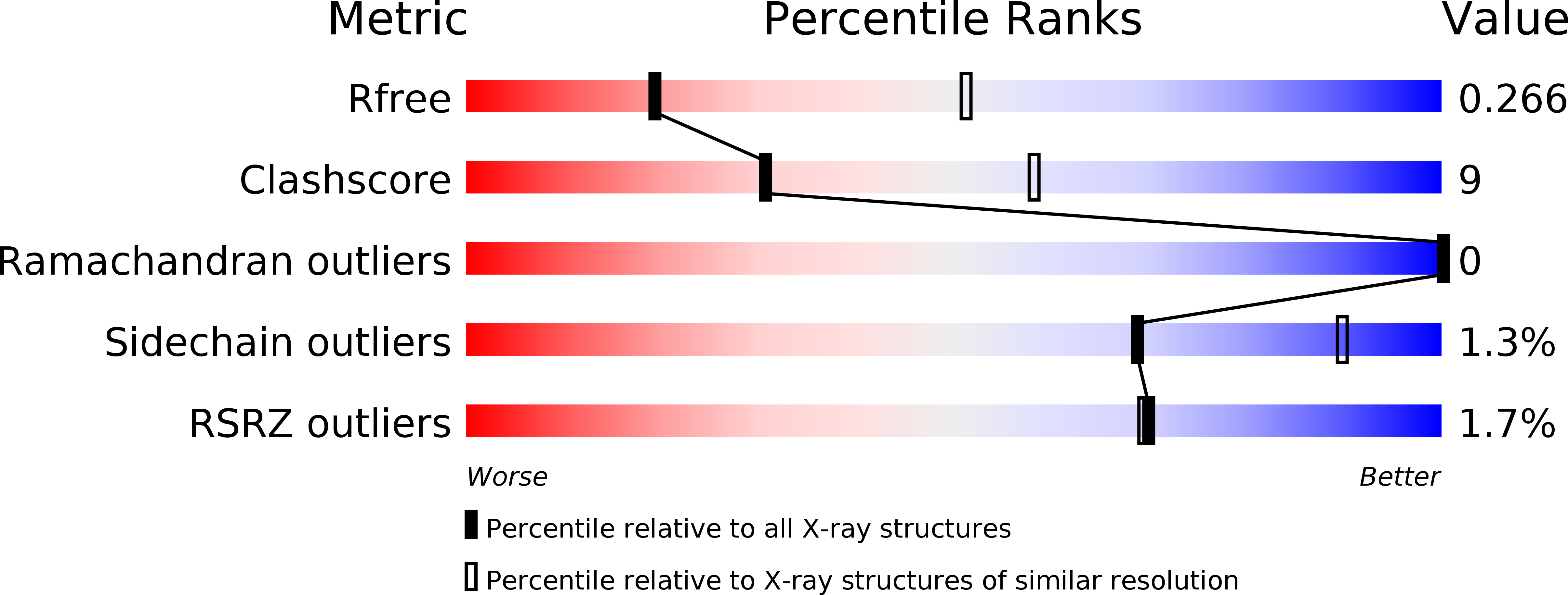
R-Value Free:
0.26
R-Value Work:
0.22
R-Value Observed:
0.23
Space Group:
P 21 21 2