6EDI image
Deposition Date 2018-08-09
Release Date 2018-08-29
Last Version Date 2023-10-11
Entry Detail
PDB ID:
6EDI
Keywords:
Title:
Crystal structure of Leishmania braziliensis glucokinase
Biological Source:
Method Details:
Experimental Method:
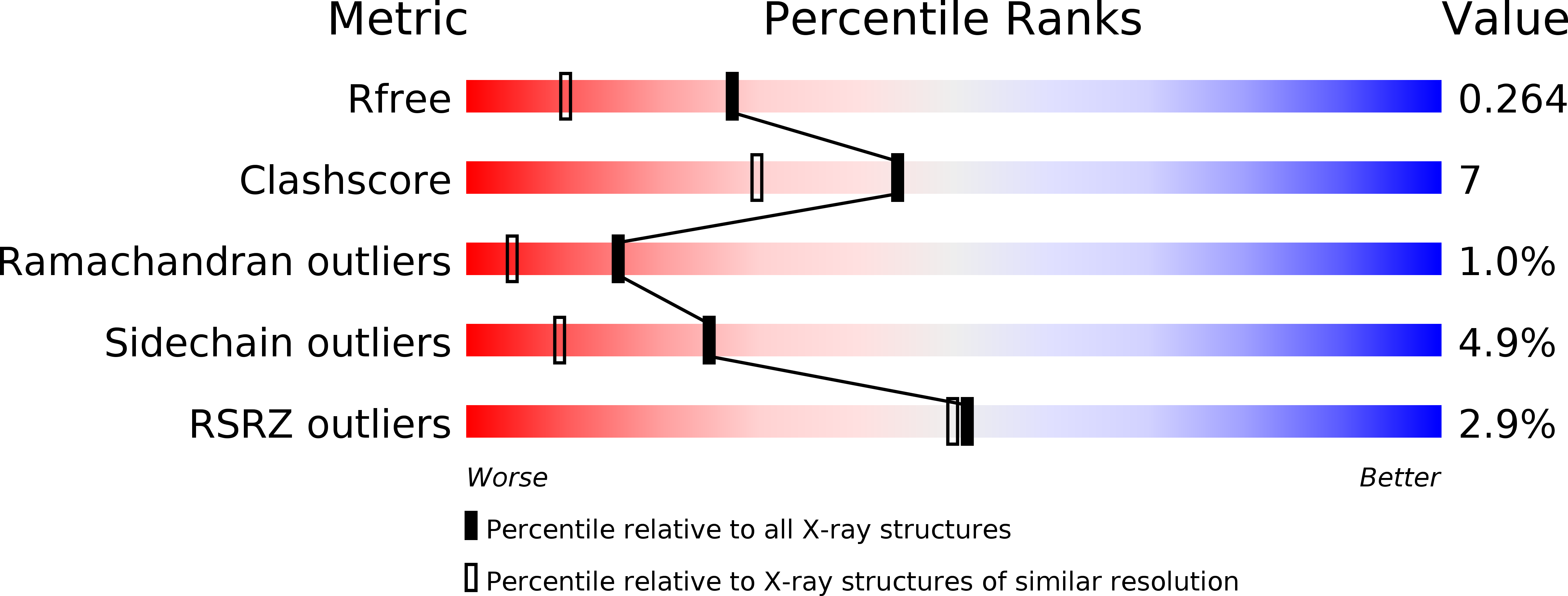
Resolution:
1.85 Å
R-Value Free:
0.26
R-Value Work:
0.21
R-Value Observed:
0.21
Space Group:
P 1 21 1
Macromolecular Entities
Structures with similar UniProt ID
Protein Blast
Polymer Type:polypeptide(L)
Molecule:Glucokinase
Gene (Uniprot):LBRM_35_2550
Chain IDs:A, B
Chain Length:412
Number of Molecules:2
Biological Source:Leishmania braziliensis MHOM/BR/75/M2904
Primary Citation
The crystal structure of glucokinase from Leishmania braziliensis.
Mol. Biochem. Parasitol. 227 47 52 (2019)
PMID: 30571993 DOI: 10.1016/j.molbiopara.2018.12.002

Abstact

Glucokinase from pathogenic protozoa of the genus Leishmania is a potential drug target for the chemotherapeutic treatment against leishmaniasis because this enzyme is located at a nodal point between two critically important metabolic pathways, glycolysis and the pentose phosphate pathway (PPP). L. braziliensis glucokinase (LbGlcK) was evaluated for its structural characterization and enzymatic performance. The enzyme catalyzes the phosphorylation of d-glucose with co-substrate ATP to yield the products G6P and ADP. LbGlcK had KM values determined as 6.61 ± 2.63 mM and 0.338 ± 0.080 mM for d-glucose and ATP, respectively. The 1.85 Å resolution X-ray crystal structure of the apo form of LbGlcK was determined and a homodimer was revealed where each subunit (both in open conformations) included the typical small and large domains. Structural comparisons were assessed in relationship to Homo sapiens hexokinase IV and Trypanosoma cruzi glucokinase. Comparisons revealed that all residues important for making hydrogen bonding interactions with d-glucose in the active site and catalysis were strictly conserved. LbGlcK was screened against four glucosamine analogue inhibitors and the stronger inhibitor of the series, HPOP-GlcN, had a Ki value of 56.9 ± 16.6 μM that exhibited competitive inhibition. For the purpose of future structure-based drug design experimentation, L. braziliensis glucokinase was observed to be very similar to T. cruzi glucokinase even though there was a 44% protein sequence identity between the two enzymes.

Legend

Protein

Chemical

Disease

Primary Citation of related structures