Deposition Date
1991-05-31
Release Date
1993-01-15
Last Version Date
2024-10-23
Entry Detail
PDB ID:
6EBX
Keywords:
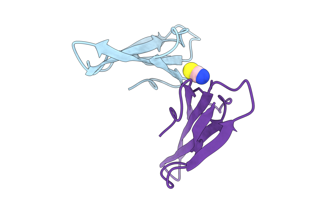
Title:
STRUCTURE DETERMINATION OF A DIMERIC FORM OF ERABUTOXIN B, CRYSTALLIZED FROM THIOCYANATE SOLUTION
Biological Source:
Source Organism(s):
Laticauda semifasciata (Taxon ID: 8631)
Method Details:
Experimental Method:
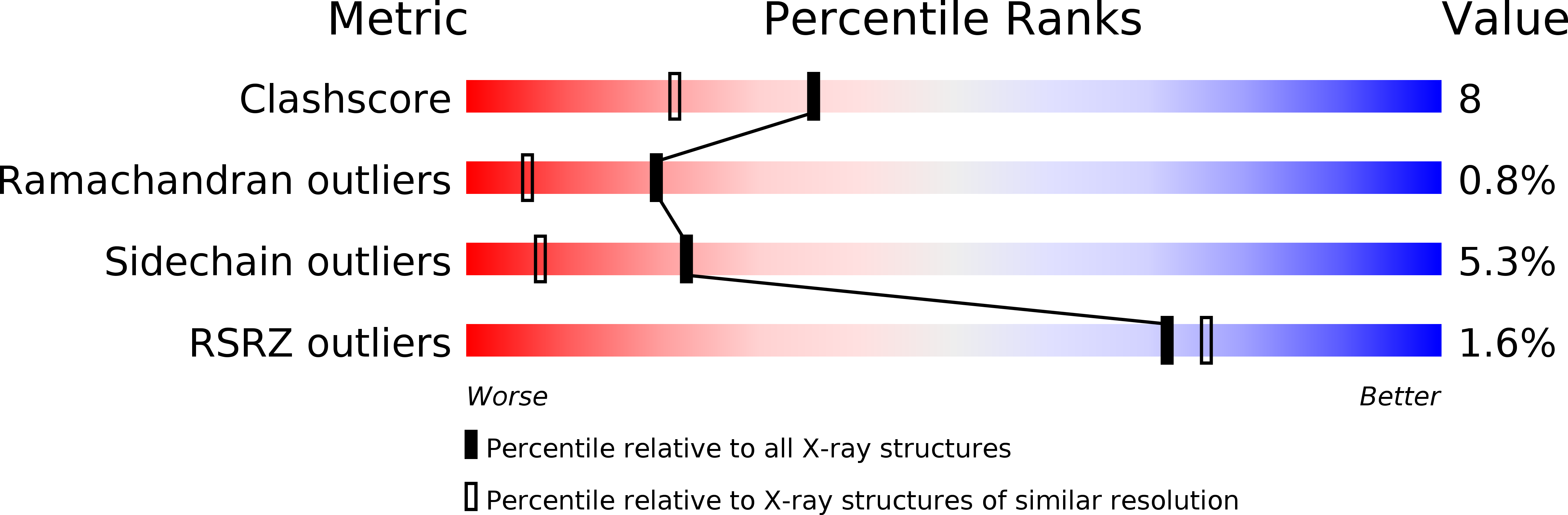
Resolution:
1.70 Å
R-Value Work:
0.19
Space Group:
P 21 21 21