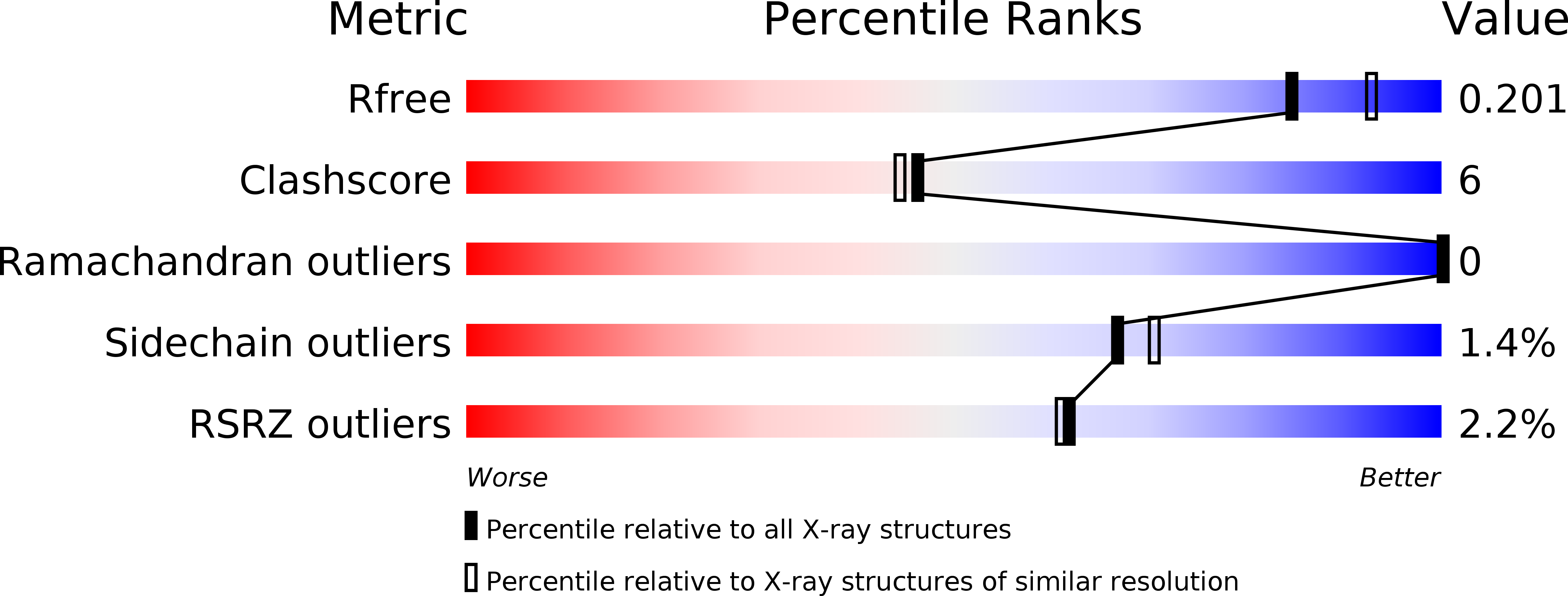
6EB6 image
Deposition Date 2018-08-05
Release Date 2019-04-10
Last Version Date 2023-10-11
Entry Detail
PDB ID:
6EB6
Keywords:
Title:
Crystal structure of BAX W139A monomer
Biological Source:
Source Organism:
Homo sapiens (Taxon ID: 9606)
Host Organism:
Method Details:
Experimental Method:
Resolution:
2.02 Å
R-Value Free:
0.20
R-Value Work:
0.17
R-Value Observed:
0.17
Space Group:
P 31 2 1
Macromolecular Entities
Polymer Type:polypeptide(L)
Molecule:Apoptosis regulator BAX
Gene (Uniprot):BAX
Mutations:W139A
Chain IDs:A
Chain Length:192
Number of Molecules:1
Biological Source:Homo sapiens
Ligand Molecules
Primary Citation
BAX Activation: Mutations Near Its Proposed Non-canonical BH3 Binding Site Reveal Allosteric Changes Controlling Mitochondrial Association.
Cell Rep 27 359 373.e6 (2019)
PMID: 30970242 DOI: 10.1016/j.celrep.2019.03.040

Abstact

To elicit apoptosis, BAX metamorphoses from an inert cytosolic monomer into homo-oligomers that permeabilize the mitochondrial outer membrane (MOM). A long-standing puzzle is that BH3 domains apparently activate BAX by not only its canonical groove but also a proposed site involving helices α1 and α6. Our mutagenesis studies reveal that late steps like oligomerization require activation through the groove but probably not earlier steps like MOM association. Conversely, α1 or α6 obstruction and alanine mutagenesis scanning implicate these helices early in BAX activation. The α1 and α6 mutations lowered BH3 binding, altered the BAX conformation, and reduced its MOM translocation and integration; their exposure of the BAX α1-α2 loop allosterically sequestered its α9 membrane anchor in the groove. The crystal structure of an α6 mutant revealed additional allosteric effects. The results suggest that the α1 and α6 region drives MOM association and integration, whereas groove binding favors subsequent steps toward oligomerization.

Legend

Protein

Chemical

Disease

Primary Citation of related structures