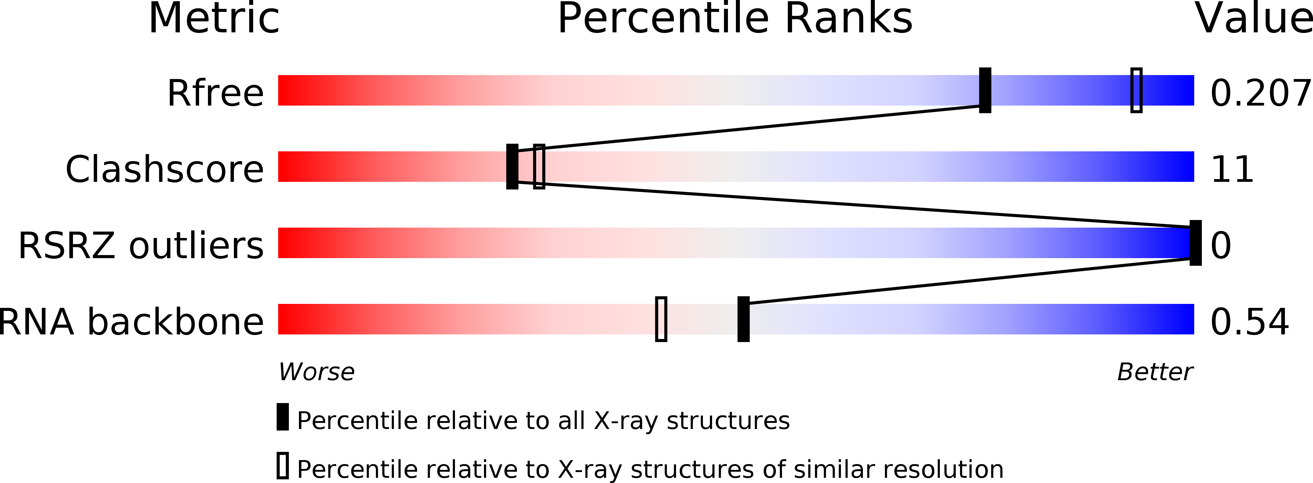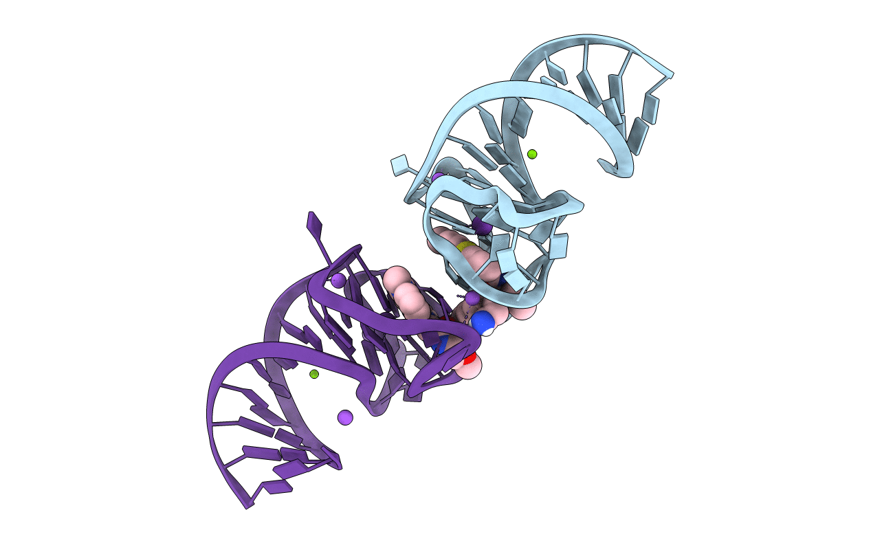
6E8S image
Deposition Date 2018-07-31
Release Date 2019-04-17
Last Version Date 2024-03-13
Entry Detail
PDB ID:
6E8S
Keywords:
Title:
Structure of the iMango-III aptamer bound to TO1-Biotin
Biological Source:
Source Organism:
Host Organism:
Method Details:
Experimental Method:
Resolution:
2.35 Å
R-Value Free:
0.20
R-Value Work:
0.18
R-Value Observed:
0.18
Space Group:
P 21 21 21
Macromolecular Entities
Polymer Type:polyribonucleotide
Molecule:iMango-III aptamer
Chain IDs:A, B
Chain Length:38
Number of Molecules:2
Biological Source:synthetic construct
Primary Citation
Structure and functional reselection of the Mango-III fluorogenic RNA aptamer.
Nat. Chem. Biol. 15 472 479 (2019)
PMID: 30992561 DOI: 10.1038/s41589-019-0267-9

Abstact

Several turn-on RNA aptamers that activate small-molecule fluorophores have been selected in vitro. Among these, the ~30 nucleotide Mango-III is notable because it binds the thiazole orange derivative TO1-Biotin with high affinity and fluoresces brightly (quantum yield 0.55). Uniquely among related aptamers, Mango-III exhibits biphasic thermal melting, characteristic of molecules with tertiary structure. We report crystal structures of TO1-Biotin complexes of Mango-III, a structure-guided mutant Mango-III(A10U), and a functionally reselected mutant iMango-III. The structures reveal a globular architecture arising from an unprecedented pseudoknot-like connectivity between a G-quadruplex and an embedded non-canonical duplex. The fluorophore is restrained into a planar conformation by the G-quadruplex, a lone, long-range trans Watson-Crick pair (whose A10U mutation increases quantum yield to 0.66), and a pyrimidine perpendicular to the nucleobase planes of those motifs. The improved iMango-III and Mango-III(A10U) fluoresce ~50% brighter than enhanced green fluorescent protein, making them suitable tags for live cell RNA visualization.

Legend

Protein

Chemical

Disease

Primary Citation of related structures