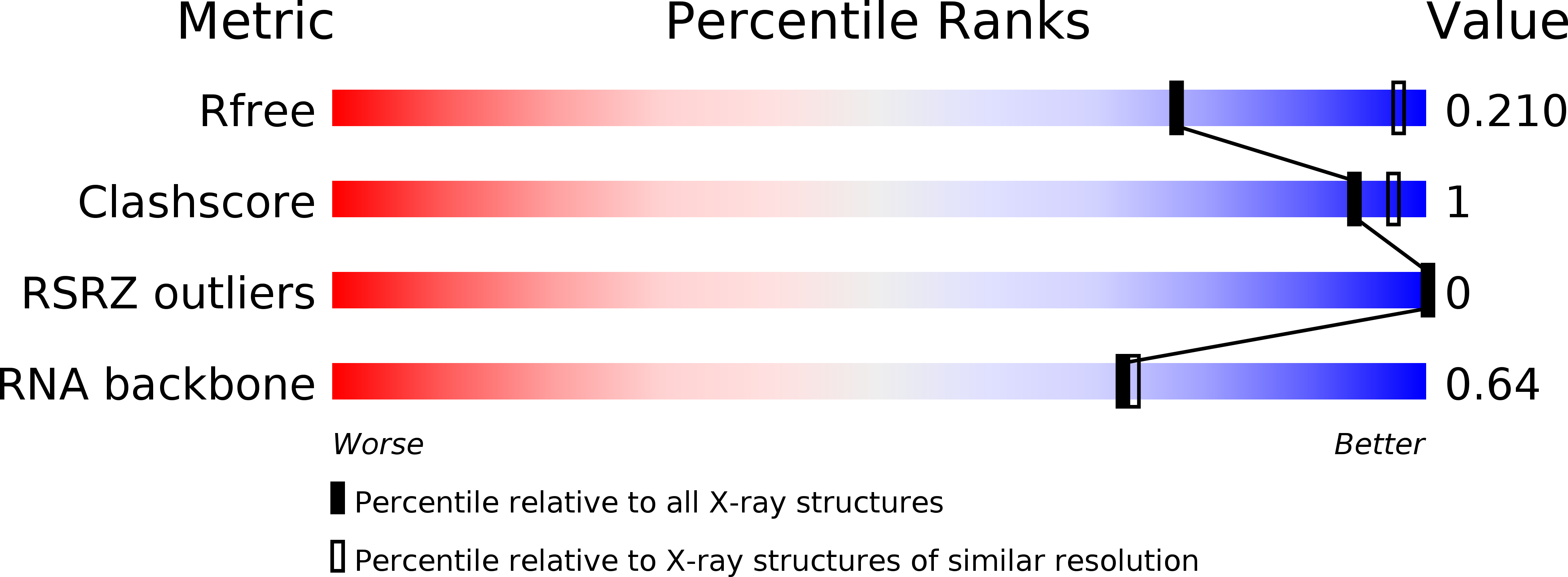
6E7L image
Deposition Date 2018-07-26
Release Date 2019-07-31
Last Version Date 2024-04-03
Entry Detail
PDB ID:
6E7L
Keywords:
Title:
Structure of a dimerized UUCG motif
Biological Source:
Source Organism:
Method Details:
Experimental Method:
Resolution:
2.59 Å
R-Value Free:
0.21
R-Value Work:
0.17
R-Value Observed:
0.17
Space Group:
P 41 21 2
Macromolecular Entities
Polymer Type:polyribonucleotide
Molecule:RNA (5'-R(*GP*GP*GP*CP*UP*GP*CP*AP*CP*UP*UP*CP*GP*GP*UP*GP*CP*UP*GP*CP*CP*C)-3')
Chain IDs:A
Chain Length:22
Number of Molecules:1
Biological Source:synthetic construct
Primary Citation
Structure of an RNA helix with pyrimidine mismatches and cross-strand stacking.
Acta Crystallogr.,Sect.F 75 652 656 (2019)
PMID: 31584014 DOI: 10.1107/S2053230X19012172

Abstact

The structure of a 22-base-pair RNA helix with mismatched pyrimidine base pairs is reported. The helix contains two symmetry-related CUG sequences: a triplet-repeat motif implicated in myotonic dystrophy type 1. The CUG repeat contains a U-U mismatch sandwiched between Watson-Crick pairs. Additionally, the center of the helix contains a dimerized UUCG motif with tandem pyrimidine (U-C/C-U) mismatches flanked by U-G wobble pairs. This region of the structure is significantly different from previously observed structures that share the same sequence and neighboring base pairs. The tandem pyrimidine mismatches are unusual and display sheared, cross-strand stacking geometries that locally constrict the helical width, a type of stacking previously associated with purines in internal loops. Thus, pyrimidine-rich regions of RNA have a high degree of structural diversity.

Legend

Protein

Chemical

Disease

Primary Citation of related structures