6E5X image
Entry Detail
PDB ID:
6E5X
Keywords:
Title:
Crystal structure of Ebola virus VP30 C-terminus/RBBP6 peptide complex
Biological Source:
PDB Version:
Deposition Date:
2018-07-23
Release Date:
2018-12-12
Method Details:
Experimental Method:
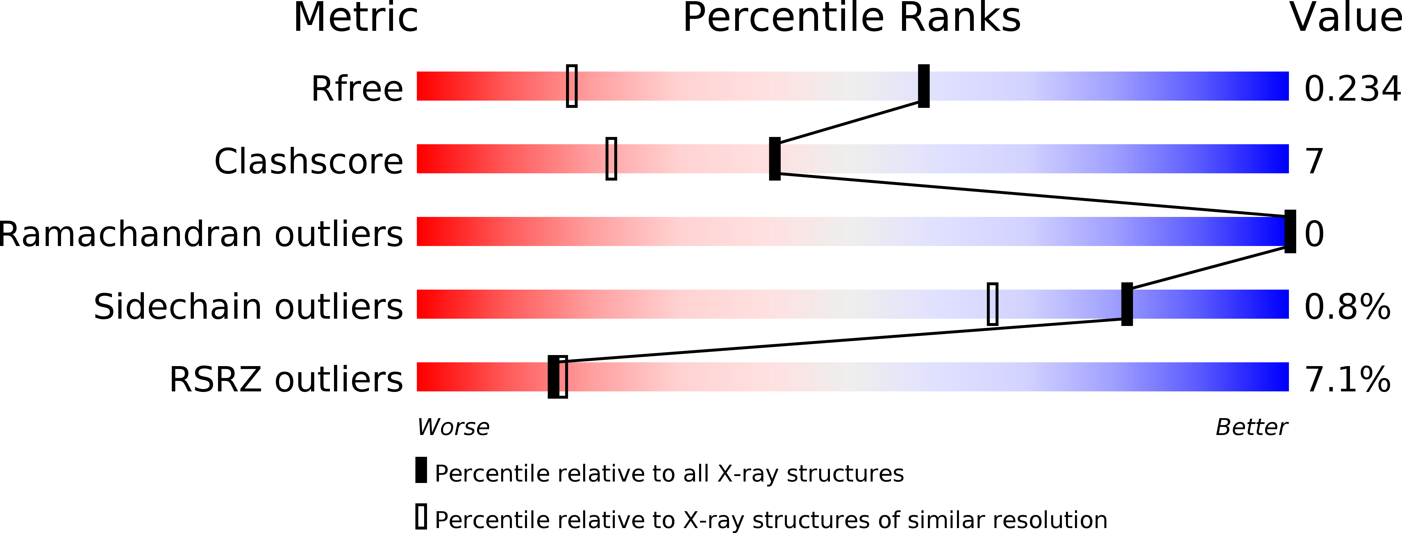
Resolution:
1.50 Å
R-Value Free:
0.23
R-Value Work:
0.18
R-Value Observed:
0.19
Space Group:
P 43 21 2
Macromolecular Entities
Polymer Type:polypeptide(L)
Description:Minor nucleoprotein VP30
Chain IDs:A
Chain Length:127
Number of Molecules:1
Biological Source:Zaire ebolavirus (strain Kikwit-95)
Polymer Type:polypeptide(L)
Description:E3 ubiquitin-protein ligase RBBP6
Chain IDs:B
Chain Length:15
Number of Molecules:1
Biological Source:Homo sapiens
Ligand Molecules
Primary Citation
Protein Interaction Mapping Identifies RBBP6 as a Negative Regulator of Ebola Virus Replication.
Cell 175 1917 1930.e13 (2018)
PMID: 30550789 DOI: 10.1016/j.cell.2018.08.044

Abstact

Ebola virus (EBOV) infection often results in fatal illness in humans, yet little is known about how EBOV usurps host pathways during infection. To address this, we used affinity tag-purification mass spectrometry (AP-MS) to generate an EBOV-host protein-protein interaction (PPI) map. We uncovered 194 high-confidence EBOV-human PPIs, including one between the viral transcription regulator VP30 and the host ubiquitin ligase RBBP6. Domain mapping identified a 23 amino acid region within RBBP6 that binds to VP30. A crystal structure of the VP30-RBBP6 peptide complex revealed that RBBP6 mimics the viral nucleoprotein (NP) binding to the same interface of VP30. Knockdown of endogenous RBBP6 stimulated viral transcription and increased EBOV replication, whereas overexpression of either RBBP6 or the peptide strongly inhibited both. These results demonstrate the therapeutic potential of biologics that target this interface and identify additional PPIs that may be leveraged for novel therapeutic strategies.

Legend

Protein

Chemical

Disease

Primary Citation of related structures