
Deposition Date
2018-07-18
Release Date
2019-04-10
Last Version Date
2023-10-11
Entry Detail
PDB ID:
6E4Y
Keywords:
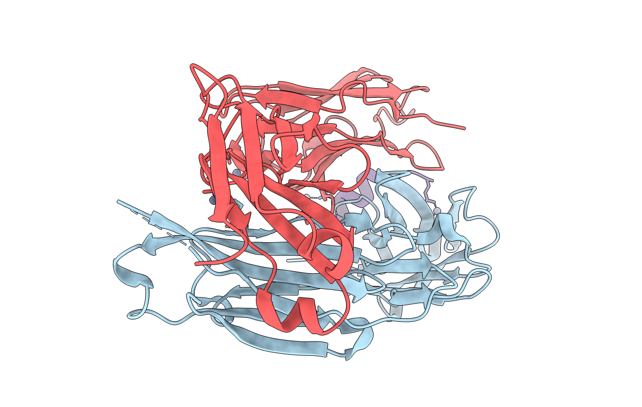
Title:
Anti-PCSK9 fab 6E2 bound to the N-terminal peptide from PCSK9, unmodified
Biological Source:
Source Organism:
Mus musculus (Taxon ID: 10090)
Homo sapiens (Taxon ID: 9606)
Homo sapiens (Taxon ID: 9606)
Host Organism:
Method Details:
Experimental Method:
Resolution:
2.24 Å
R-Value Free:
0.25
R-Value Work:
0.21
R-Value Observed:
0.21
Space Group:
P 21 21 2
