6E2J image
Deposition Date 2018-07-11
Release Date 2019-05-15
Last Version Date 2024-03-13
Entry Detail
PDB ID:
6E2J
Keywords:
Title:
Crystal structure of the heterocomplex between human keratin 1 coil 1B containing S233L mutation and wild-type human keratin 10 coil 1B
Biological Source:
Source Organism:
Homo sapiens (Taxon ID: 9606)
Host Organism:
Method Details:
Experimental Method:
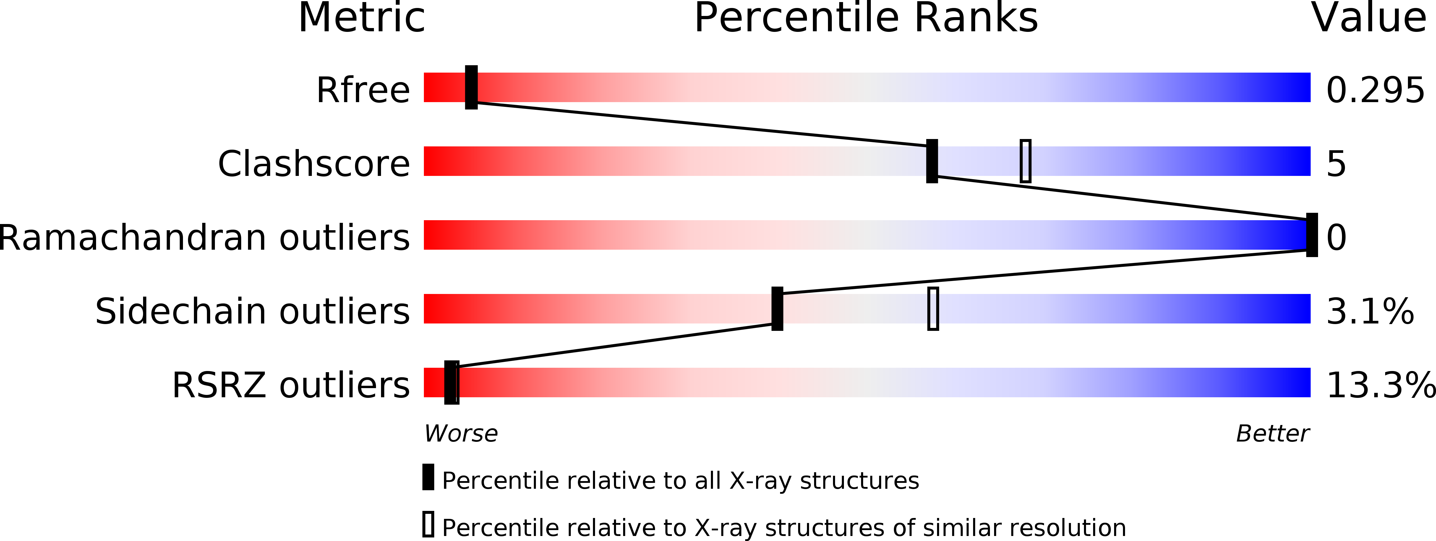
Resolution:
2.39 Å
R-Value Free:
0.29
R-Value Work:
0.27
R-Value Observed:
0.27
Space Group:
P 64 2 2
Macromolecular Entities
Structures with similar UniProt ID
Protein Blast
Polymer Type:polypeptide(L)
Molecule:Keratin, type II cytoskeletal 1
Gene (Uniprot):KRT1
Mutagens:S233L
Chain IDs:A
Chain Length:107
Number of Molecules:1
Biological Source:Homo sapiens
Structures with similar UniProt ID
Protein Blast
Polymer Type:polypeptide(L)
Molecule:Keratin, type I cytoskeletal 10
Gene (Uniprot):KRT10
Chain IDs:B
Chain Length:104
Number of Molecules:1
Biological Source:Homo sapiens
Ligand Molecules
Primary Citation
Human keratin 1/10-1B tetramer structures reveal a knob-pocket mechanism in intermediate filament assembly.
Embo J. 38 ? ? (2019)
PMID: 31036554 DOI: 10.15252/embj.2018100741

Abstact

To characterize keratin intermediate filament assembly mechanisms at atomic resolution, we determined the crystal structure of wild-type human keratin-1/keratin-10 helix 1B heterotetramer at 3.0 Å resolution. It revealed biochemical determinants for the A11 mode of axial alignment in keratin filaments. Four regions on a hydrophobic face of the K1/K10-1B heterodimer dictated tetramer assembly: the N-terminal hydrophobic pocket (defined by L227K1, Y230K1, F231K1, and F234K1), the K10 hydrophobic stripe, K1 interaction residues, and the C-terminal anchoring knob (formed by F314K1 and L318K1). Mutation of both knob residues to alanine disrupted keratin 1B tetramer and full-length filament assembly. Individual knob residue mutant F314AK1, but not L318AK1, abolished 1B tetramer formation. The K1-1B knob/pocket mechanism is conserved across keratins and many non-keratin intermediate filaments. To demonstrate how pathogenic mutations cause skin disease by altering filament assembly, we additionally determined the 2.39 Å structure of K1/10-1B containing a S233LK1 mutation linked to epidermolytic palmoplantar keratoderma. Light scattering and circular dichroism measurements demonstrated enhanced aggregation of K1S233L/K10-1B in solution without affecting secondary structure. The K1S233L/K10-1B octamer structure revealed S233LK1 causes aberrant hydrophobic interactions between 1B tetramers.

Legend

Protein

Chemical

Disease

Primary Citation of related structures