6E2A image
Deposition Date 2018-07-10
Release Date 2019-02-06
Last Version Date 2023-10-11
Entry Detail
PDB ID:
6E2A
Title:
Crystal structure of NADH:quinone reductase PA1024 from Pseudomonas aeruginosa PAO1 in complex with NAD+
Biological Source:
Method Details:
Experimental Method:
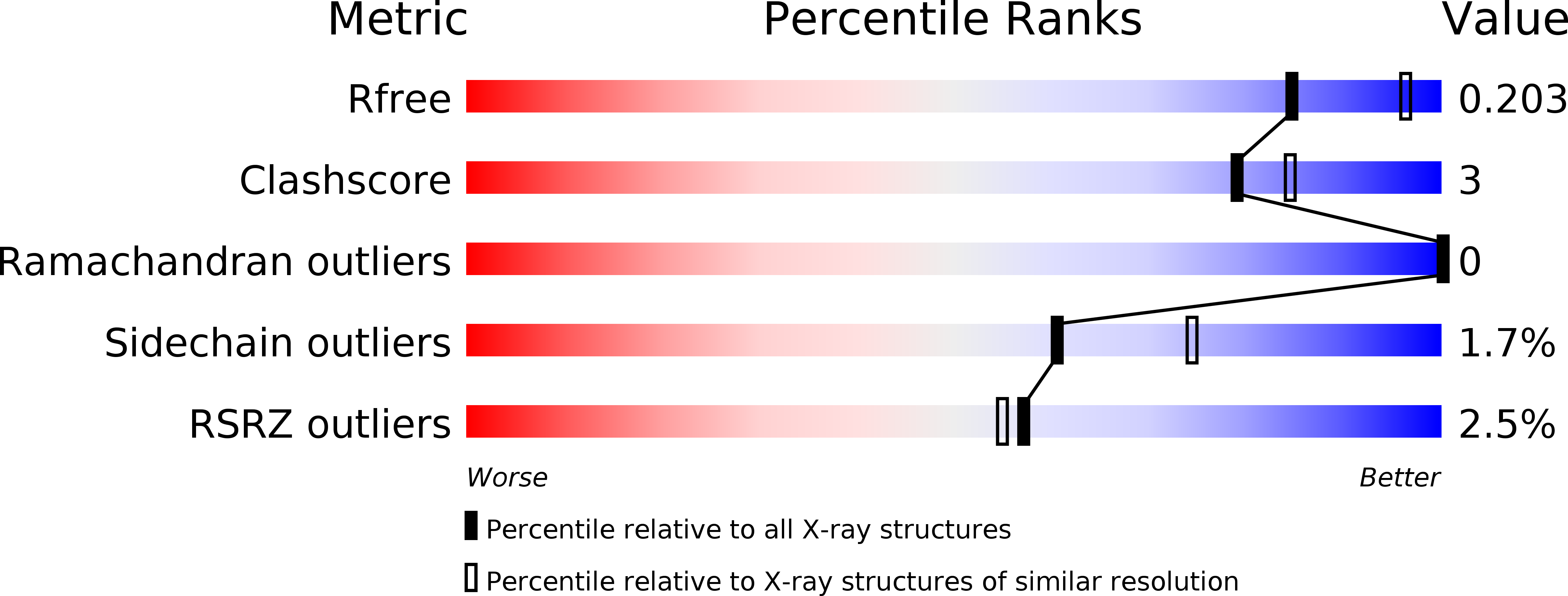
Resolution:
2.20 Å
R-Value Free:
0.20
R-Value Work:
0.15
R-Value Observed:
0.15
Space Group:
P 31 2 1
Macromolecular Entities
Polymer Type:polypeptide(L)
Molecule:Nitronate monooxygenase
Gene (Uniprot):PA1024
Chain IDs:A
Chain Length:334
Number of Molecules:1
Biological Source:Pseudomonas aeruginosa (strain ATCC 15692 / DSM 22644 / CIP 104116 / JCM 14847 / LMG 12228 / 1C / PRS 101 / PAO1)
Primary Citation
Steric hindrance controls pyridine nucleotide specificity of a flavin-dependent NADH:quinone oxidoreductase.
Protein Sci. 28 167 175 (2019)
PMID: 30246917 DOI: 10.1002/pro.3514

Abstact

The crystal structure of the NADH:quinone oxidoreductase PA1024 has been solved in complex with NAD+ to 2.2 Å resolution. The nicotinamide C4 is 3.6 Å from the FMN N5 atom, with a suitable orientation for facile hydride transfer. NAD+ binds in a folded conformation at the interface of the TIM-barrel domain and the extended domain of the enzyme. Comparison of the enzyme-NAD+ structure with that of the ligand-free enzyme revealed a different conformation of a short loop (75-86) that is part of the NAD+ -binding pocket. P78, P82, and P84 provide internal rigidity to the loop, whereas Q80 serves as an active site latch that secures the NAD+ within the binding pocket. An interrupted helix consisting of two α-helices connected by a small three-residue loop binds the pyrophosphate moiety of NAD+ . The adenine moiety of NAD+ appears to π-π stack with Y261. Steric constraints between the adenosine ribose of NAD+ , P78, and Q80, control the strict specificity of the enzyme for NADH. Charged residues do not play a role in the specificity of PA1024 for the NADH substrate.

Legend

Protein

Chemical

Disease

Primary Citation of related structures