
Deposition Date
2018-06-28
Release Date
2018-09-19
Last Version Date
2023-10-11
Entry Detail
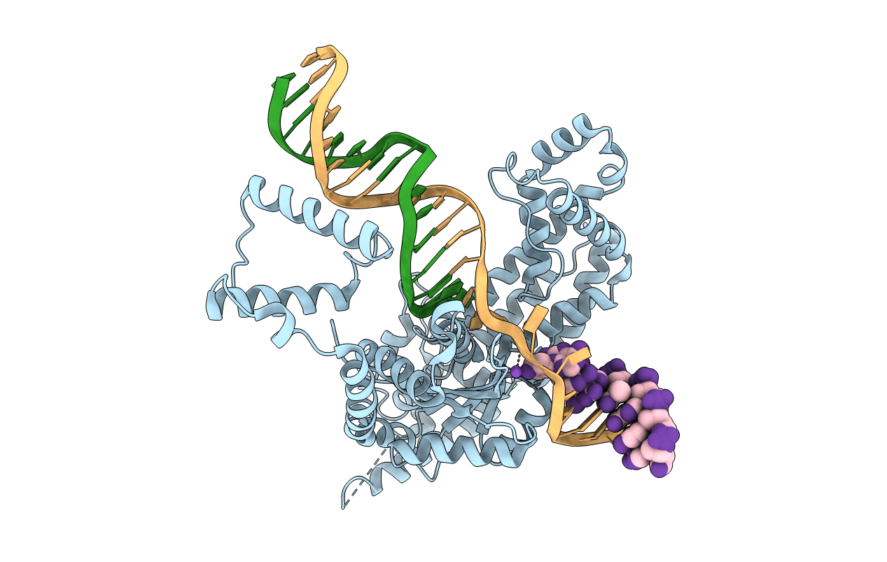
PDB ID:
6DX0
Keywords:
Title:
Hermes transposase deletion dimer complex with (A/T) DNA
Biological Source:
Source Organism(s):
Musca domestica (Taxon ID: 7370)
Expression System(s):
Method Details:
Experimental Method:
Resolution:
2.90 Å
R-Value Free:
0.30
R-Value Work:
0.22
R-Value Observed:
0.22
Space Group:
C 2 2 21
