
Deposition Date
2018-06-11
Release Date
2019-01-09
Last Version Date
2023-10-11
Entry Detail
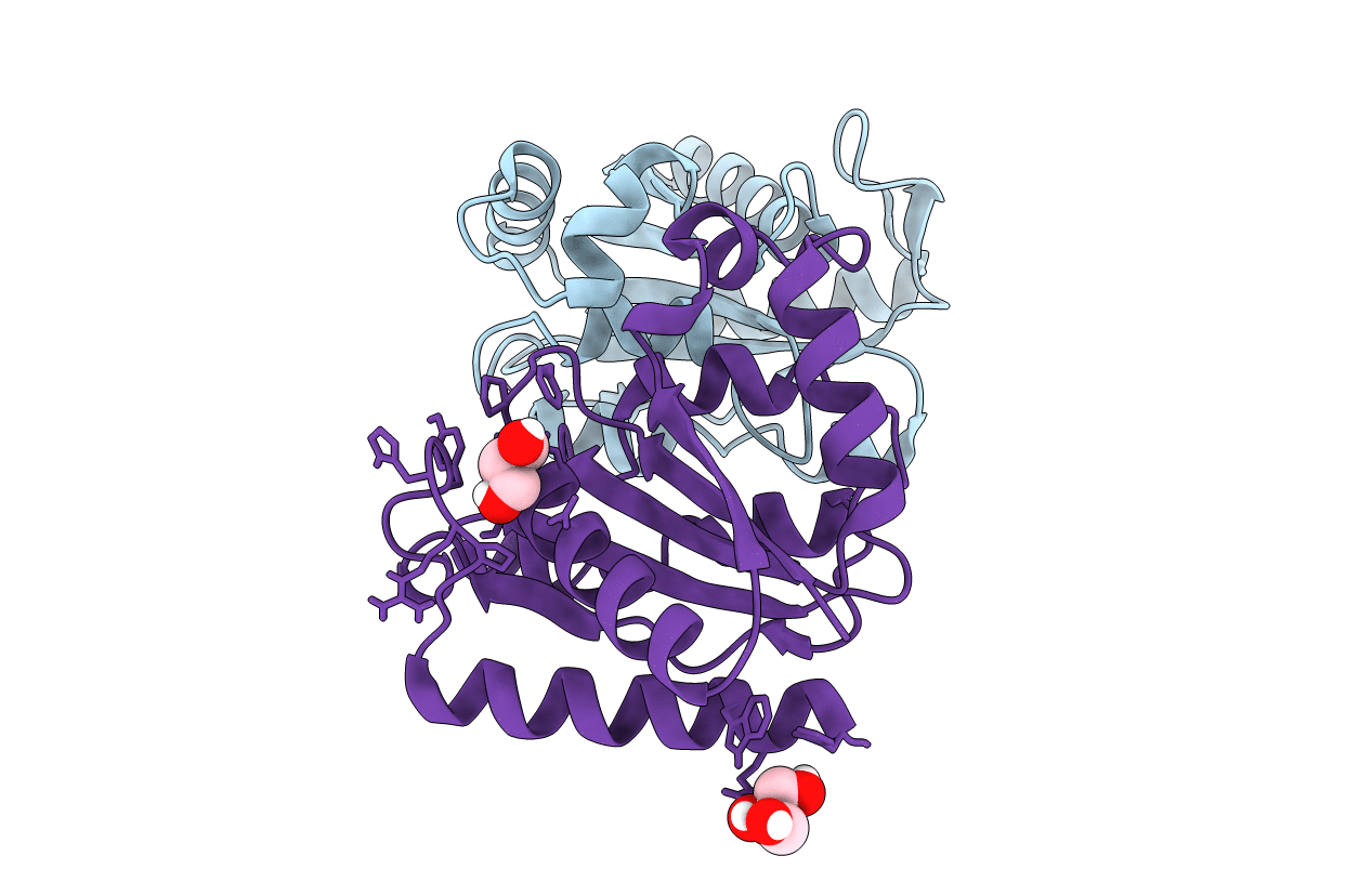
PDB ID:
6DQP
Keywords:
Title:
Crystal structure of SsuE FMN reductase Delta118 mutant in apo form
Biological Source:
Source Organism(s):
Escherichia coli (strain K12) (Taxon ID: 83333)
Expression System(s):
Method Details:
Experimental Method:
Resolution:
1.55 Å
R-Value Free:
0.17
R-Value Work:
0.16
R-Value Observed:
0.16
Space Group:
P 21 21 21
