Deposition Date
2018-05-30
Release Date
2018-07-18
Last Version Date
2023-10-11
Entry Detail
PDB ID:
6DKU
Keywords:
Title:
Crystal structure of Myotis VP35 mutant of interferon inhibitory domain
Biological Source:
Source Organism:
Myotis lucifugus (Taxon ID: 59463)
Host Organism:
Method Details:
Experimental Method:
Resolution:
2.60 Å
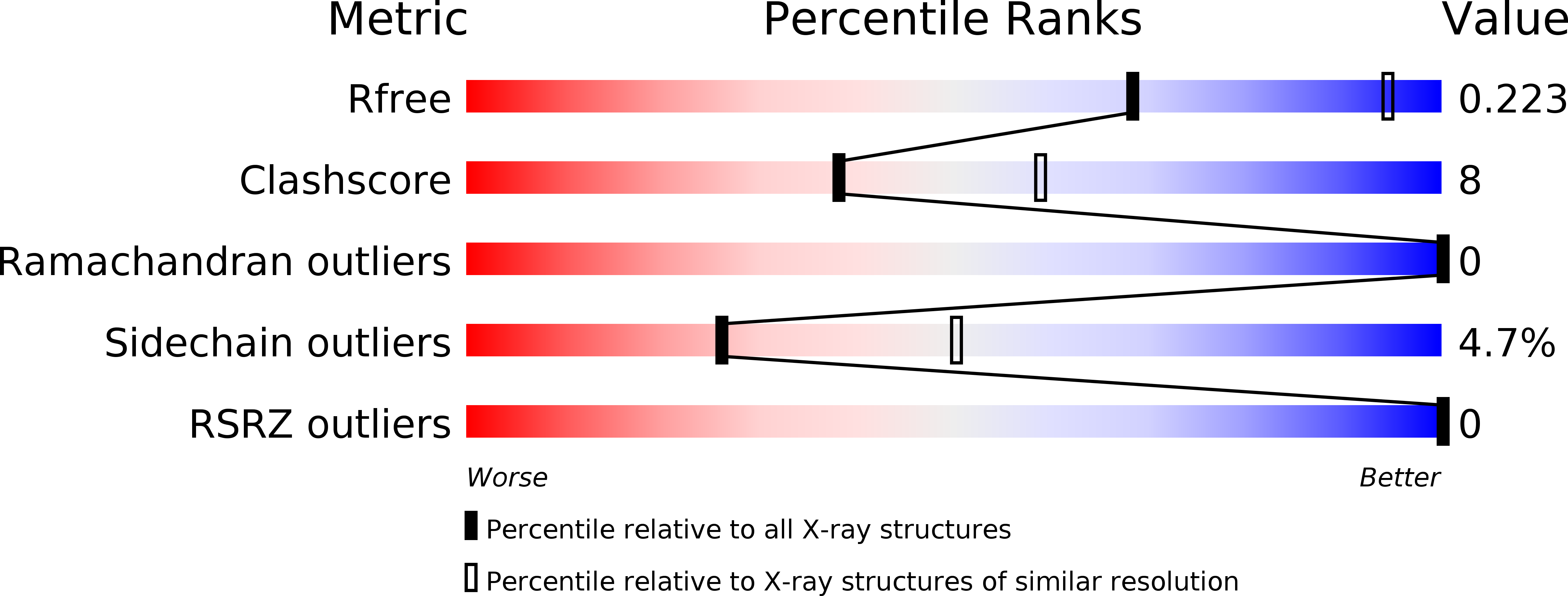
R-Value Free:
0.22
R-Value Work:
0.17
R-Value Observed:
0.17
Space Group:
P 32 2 1