6DJW image
Deposition Date 2018-05-26
Release Date 2018-07-04
Last Version Date 2024-10-30
Entry Detail
PDB ID:
6DJW
Keywords:
Title:
Crystal Structure of pParkin (REP and RING2 deleted)-pUb-UbcH7 complex
Biological Source:
Source Organism:
Bactrocera dorsalis (Taxon ID: 27457)
Homo sapiens (Taxon ID: 9606)
Bos taurus (Taxon ID: 9913)
Method Details:
Experimental Method:
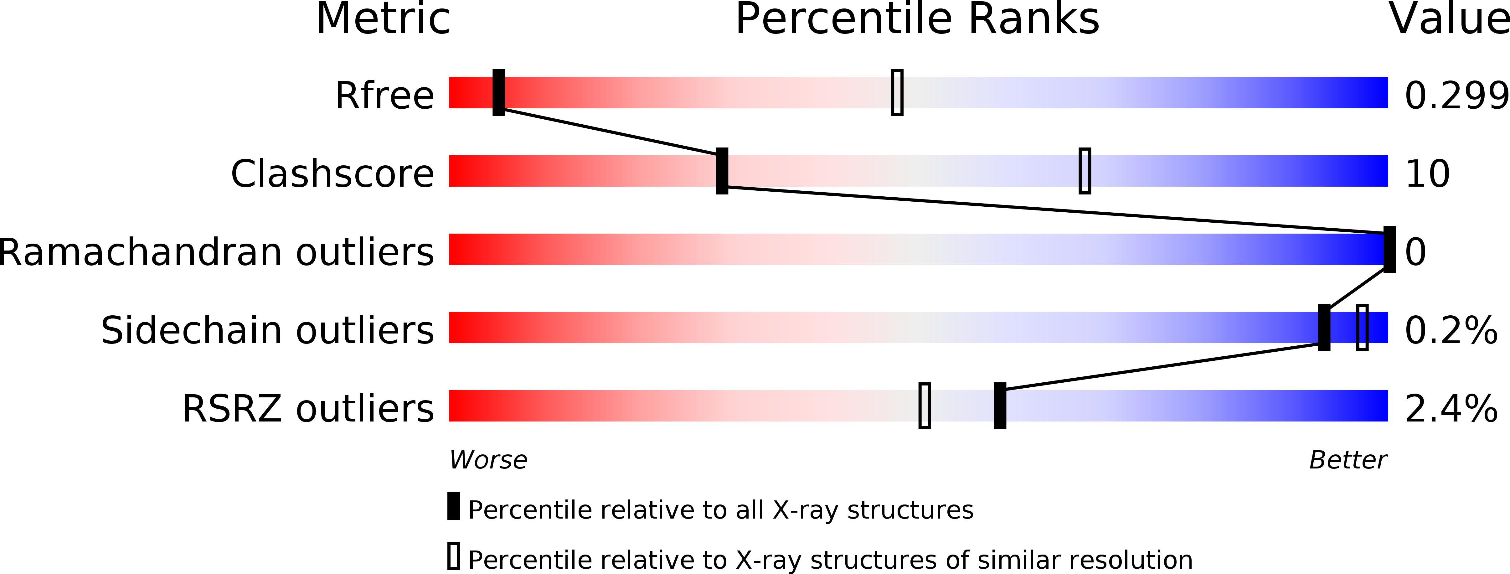
Resolution:
3.80 Å
R-Value Free:
0.29
R-Value Work:
0.26
R-Value Observed:
0.26
Space Group:
P 31 2 1
Macromolecular Entities
Structures with similar UniProt ID
Protein Blast
Polymer Type:polypeptide(L)
Molecule:RBR-type E3 ubiquitin transferase,RBR-type E3 ubiquitin transferase
Gene (Uniprot):PRKN2
Chain IDs:A
Chain Length:349
Number of Molecules:1
Biological Source:Bactrocera dorsalis
Structures with similar UniProt ID
Protein Blast
Polymer Type:polypeptide(L)
Molecule:Ubiquitin
Gene (Uniprot):RPS27A
Chain IDs:B
Chain Length:74
Number of Molecules:1
Biological Source:Bos taurus
Structures with similar UniProt ID
Protein Blast
Polymer Type:polypeptide(L)
Molecule:Ubiquitin-conjugating enzyme E2 L3
Gene (Uniprot):UBE2L3
Mutagens:C86K
Chain IDs:C
Chain Length:159
Number of Molecules:1
Biological Source:Homo sapiens
Modified Residue
Compound ID Chain ID Parent Comp ID Details 2D Image
SEP A SER modified residue
Ligand Molecules
Primary Citation
Mechanism of parkin activation by phosphorylation.
Nat. Struct. Mol. Biol. 25 623 630 (2018)
PMID: 29967542 DOI: 10.1038/s41594-018-0088-7

Abstact

Mutations in the ubiquitin ligase parkin are responsible for a familial form of Parkinson's disease. Parkin and the PINK1 kinase regulate a quality-control system for mitochondria. PINK1 phosphorylates ubiquitin on the outer membrane of damaged mitochondria, thus leading to recruitment and activation of parkin via phosphorylation of its ubiquitin-like (Ubl) domain. Here, we describe the mechanism of parkin activation by phosphorylation. The crystal structure of phosphorylated Bactrocera dorsalis (oriental fruit fly) parkin in complex with phosphorylated ubiquitin and an E2 ubiquitin-conjugating enzyme reveals that the key activating step is movement of the Ubl domain and release of the catalytic RING2 domain. Hydrogen/deuterium exchange and NMR experiments with the various intermediates in the activation pathway confirm and extend the interpretation of the crystal structure to mammalian parkin. Our results rationalize previously unexplained Parkinson's disease mutations and the presence of internal linkers that allow large domain movements in parkin.

Legend

Protein

Chemical

Disease

Primary Citation of related structures