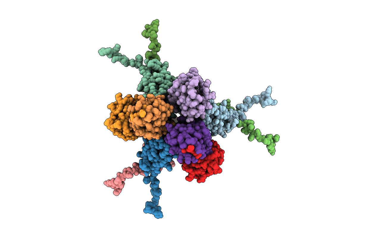
6DJ9 image
Entry Detail
PDB ID:
6DJ9
Title:
Structure of the USP15 DUSP domain in complex with a high-affinity Ubiquitin Variant (UbV)
Biological Source:
Source Organism:
Host Organism:
PDB Version:
Deposition Date:
2018-05-24
Release Date:
2019-01-23
Method Details:
Experimental Method:
Resolution:
3.10 Å
R-Value Free:
0.26
R-Value Work:
0.25
R-Value Observed:
0.25
Space Group:
P 63
Macromolecular Entities
Polymer Type:polypeptide(L)
Description:Ubiquitin carboxyl-terminal hydrolase 15
Chain IDs:A, B, C, D, E, F
Chain Length:141
Number of Molecules:6
Biological Source:Homo sapiens
Polymer Type:polypeptide(L)
Description:Ubiquitin Variant UbV 15.D
Chain IDs:G, H, I, J, K, L
Chain Length:86
Number of Molecules:6
Biological Source:Homo sapiens
Ligand Molecules
Primary Citation

Abstact

The multi-domain deubiquitinase USP15 regulates diverse eukaryotic processes and has been implicated in numerous diseases. We developed ubiquitin variants (UbVs) that targeted either the catalytic domain or each of three adaptor domains in USP15, including the N-terminal DUSP domain. We also designed a linear dimer (diUbV), which targeted the DUSP and catalytic domains, and exhibited enhanced specificity and more potent inhibition of catalytic activity than either UbV alone. In cells, the UbVs inhibited the deubiquitination of two USP15 substrates, SMURF2 and TRIM25, and the diUbV inhibited the effects of USP15 on the transforming growth factor β pathway. Structural analyses revealed that three distinct UbVs bound to the catalytic domain and locked the active site in a closed, inactive conformation, and one UbV formed an unusual strand-swapped dimer and bound two DUSP domains simultaneously. These inhibitors will enable the study of USP15 function in oncology, neurology, immunology, and inflammation.

Legend

Protein

Chemical

Disease

Primary Citation of related structures