6DJ0 image
Deposition Date 2018-05-24
Release Date 2018-10-31
Last Version Date 2024-04-03
Entry Detail
PDB ID:
6DJ0
Keywords:
Title:
ASLTVS segment from Human Immunoglobulin Light-Chain Variable Domain, Residues 73-78, assembled as an amyloid fibril
Biological Source:
Source Organism:
Homo sapiens (Taxon ID: 9606)
Method Details:
Experimental Method:
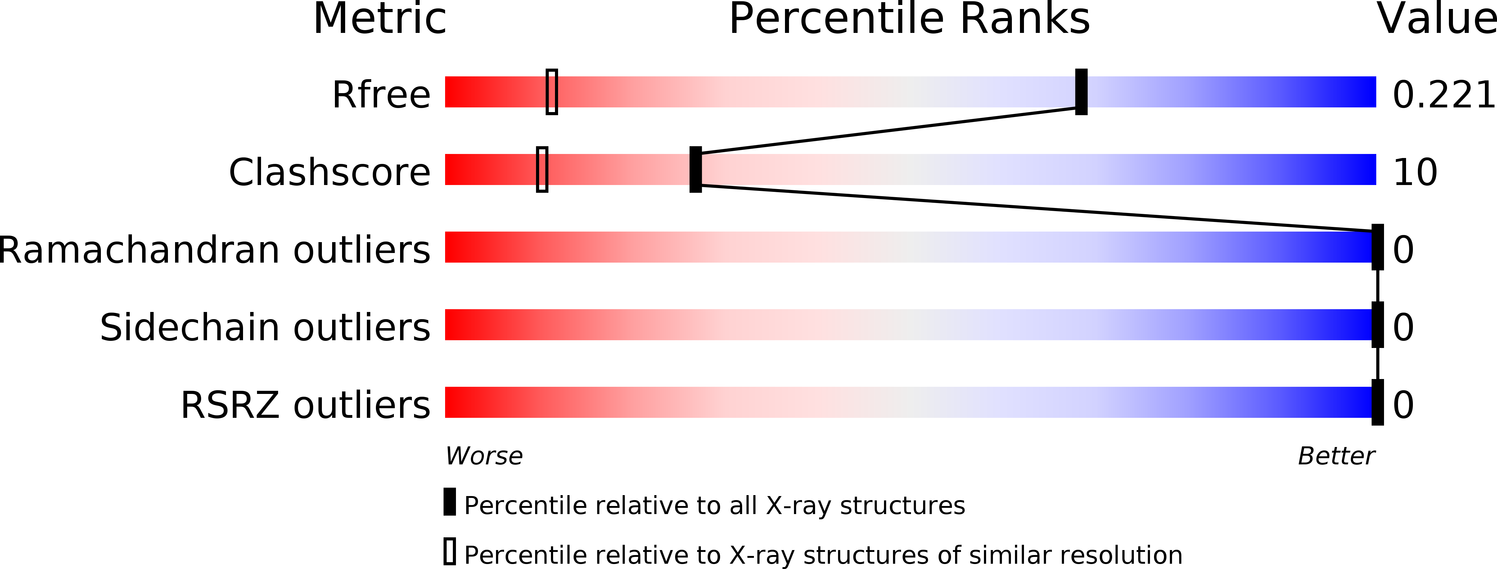
Resolution:
1.30 Å
R-Value Free:
0.21
R-Value Work:
0.18
R-Value Observed:
0.18
Space Group:
P 1 21 1
Macromolecular Entities
Polymer Type:polypeptide(L)
Molecule:ASLTVS segment from Light-Chain Variable Domain, Lambda Mcg
Chain IDs:A, B
Chain Length:6
Number of Molecules:2
Biological Source:Homo sapiens
Primary Citation
Identification of two principal amyloid-driving segments in variable domains of Ig light chains in systemic light-chain amyloidosis.
J. Biol. Chem. 293 19659 19671 (2018)
PMID: 30355736 DOI: 10.1074/jbc.RA118.004142

Abstact

Systemic light-chain amyloidosis (AL) is a human disease caused by overexpression of monoclonal immunoglobulin light chains that form pathogenic amyloid fibrils. These amyloid fibrils deposit in tissues and cause organ failure. Proteins form amyloid fibrils when they partly or fully unfold and expose segments capable of stacking into β-sheets that pair and thereby form a tight, dehydrated interface. These structures, termed steric zippers, constitute the spines of amyloid fibrils. Here, using a combination of computational (with ZipperDB and Boston University ALBase), mutational, biochemical, and protein structural analyses, we identified segments within the variable domains of Ig light chains that drive the assembly of amyloid fibrils in AL. We demonstrate that there are at least two such segments and that each one can drive amyloid fibril assembly independently of the other. Our analysis revealed that peptides derived from these segments form steric zippers featuring a typical dry interface with high-surface complementarity and occupy the same spatial location of the Greek-key immunoglobulin fold in both λ and κ variable domains. Of note, some predicted steric-zipper segments did not form amyloid fibrils or assembled into fibrils only when removed from the whole protein. We conclude that steric-zipper propensity must be experimentally validated and that the two segments identified here may represent therapeutic targets. In addition to elucidating the molecular pathogenesis of AL, these findings also provide an experimental approach for identifying segments that drive fibril formation in other amyloid diseases.

Legend

Protein

Chemical

Disease

Primary Citation of related structures