Deposition Date
2018-05-23
Release Date
2018-09-05
Last Version Date
2024-11-20
Entry Detail
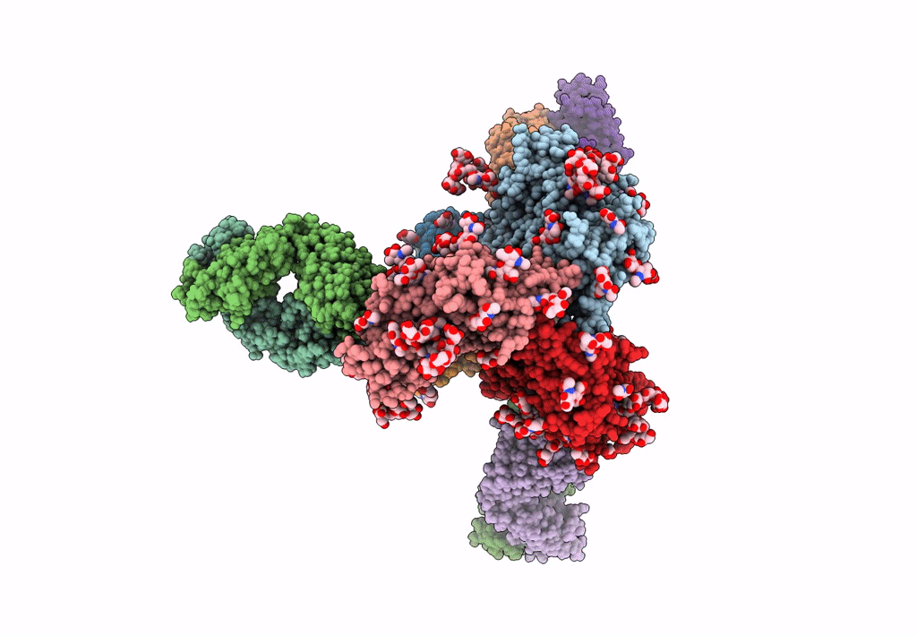
PDB ID:
6DID
Keywords:
Title:
HIV Env BG505 SOSIP with polyclonal Fabs from immunized rabbit #3417 post-boost#1
Biological Source:
Source Organism(s):
Human immunodeficiency virus 1 (Taxon ID: 11676)
Oryctolagus cuniculus (Taxon ID: 9986)
Oryctolagus cuniculus (Taxon ID: 9986)
Expression System(s):
Method Details:
Experimental Method:
Resolution:
4.71 Å
Aggregation State:
PARTICLE
Reconstruction Method:
SINGLE PARTICLE