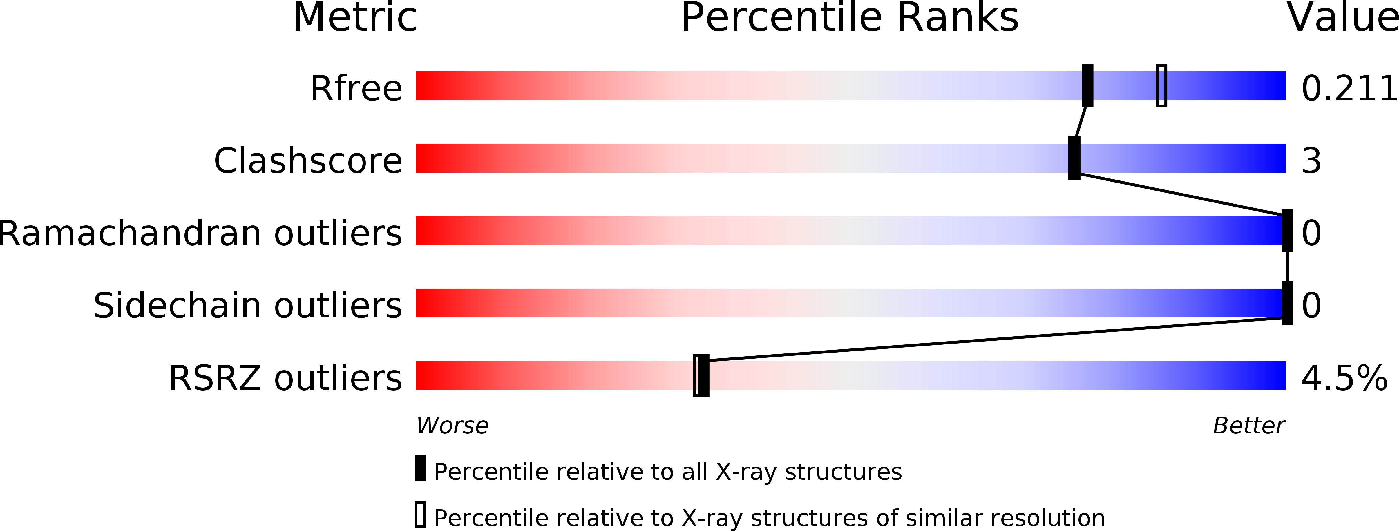
6DHW image
Deposition Date 2018-05-21
Release Date 2018-12-19
Last Version Date 2024-11-06
Entry Detail
PDB ID:
6DHW
Keywords:
Title:
Crystal structure of primase iron-sulfur domain (266-457)
Biological Source:
Source Organism:
Homo sapiens (Taxon ID: 9606)
Host Organism:
Method Details:
Experimental Method:
Resolution:
2.01 Å
R-Value Free:
0.21
R-Value Work:
0.18
R-Value Observed:
0.18
Space Group:
C 1 2 1
Macromolecular Entities
Structures with similar UniProt ID
Protein Blast
Polymer Type:polypeptide(L)
Molecule:DNA primase large subunit
Gene (Uniprot):PRIM2
Chain IDs:A, B
Chain Length:192
Number of Molecules:2
Biological Source:Homo sapiens
Primary Citation
Functional and structural similarity of human DNA primase [4Fe4S] cluster domain constructs.
PLoS ONE 13 e0209345 e0209345 (2018)
PMID: 30562384 DOI: 10.1371/journal.pone.0209345

Abstact

The regulatory subunit of human DNA primase has a C-terminal domain (p58C) that contains a [4Fe4S] cluster and binds DNA. Previous electrochemical analysis of a p58C construct revealed that its affinity for DNA is sensitive to the redox state of the [4Fe4S] cluster. Concerns about the validity of this conclusion have been raised, based in part on differences in X-ray crystal structures of the p58C272-464 construct used for that study and that of a N-terminally shifted p58C266-456 construct and consequently, an assumption that p58C272-464 has abnormal physical and functional properties. To address this controversy, a new p58C266-464 construct containing all residues was crystallized under the conditions previously used for crystallizing p58C272-464, and the solution structures of both constructs were assessed using circular dichroism and NMR spectroscopy. In the new crystal structure, p58C266-464 exhibits the same elements of secondary structure near the DNA binding site as observed in the crystal structure of p58C272-464. Moreover, in solution, circular dichroism and 15N,1H-heteronuclear single quantum coherence (HSQC) NMR spectra show there are no significant differences in the distribution of secondary structures or in the tertiary structure or the two constructs. To validate that the two constructs have the same functional properties, binding of a primed DNA template was measured using a fluorescence-based DNA binding assay, and the affinities for this substrate were the same (3.4 ± 0.5 μM and 2.7 ± 0.3 μM, respectively). The electrochemical properties of p58C266-464 were also measured and this p58C construct was able to engage in redox switching on DNA with the same efficiency as p58C272-464. Together, these results show that although p58C can be stabilized in different conformations in the crystalline state, in solution there is effectively no difference in the structure and functional properties of p58C constructs of different lengths.

Legend

Protein

Chemical

Disease

Primary Citation of related structures