6DFX image
Entry Detail
PDB ID:
6DFX
Keywords:
Title:
human diabetogenic TCR T1D3 in complex with DQ8-p8E9E peptide
Biological Source:
Source Organism:
Host Organism:
PDB Version:
Deposition Date:
2018-05-15
Release Date:
2019-04-17
Method Details:
Experimental Method:
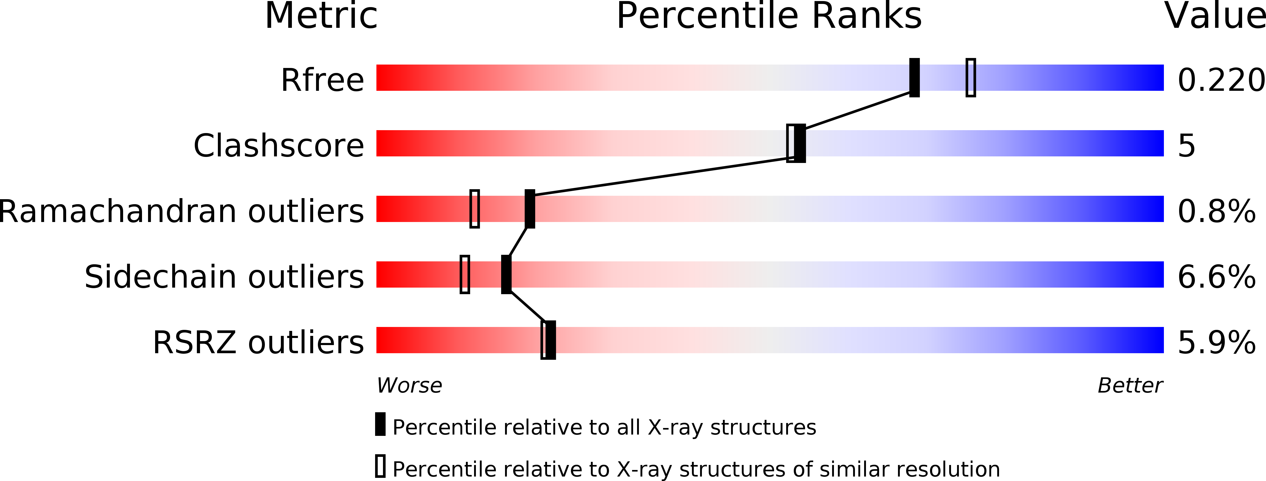
Resolution:
2.03 Å
R-Value Free:
0.21
R-Value Work:
0.18
R-Value Observed:
0.18
Space Group:
P 1 21 1
Macromolecular Entities
Polymer Type:polypeptide(L)
Description:MHC class II HLA-DQ-alpha chain
Chain IDs:A, C (auth: D)
Chain Length:188
Number of Molecules:2
Biological Source:Homo sapiens
Polymer Type:polypeptide(L)
Description:MHC class II antigen
Chain IDs:B, D (auth: E)
Chain Length:221
Number of Molecules:2
Biological Source:Homo sapiens
Polymer Type:polypeptide(L)
Description:T1D3 alpha chain
Chain IDs:E (auth: G), G (auth: I)
Chain Length:207
Number of Molecules:2
Biological Source:Homo sapiens
Polymer Type:polypeptide(L)
Description:T1D3 beta chain
Chain IDs:F (auth: H), H (auth: J)
Chain Length:238
Number of Molecules:2
Biological Source:Homo sapiens
Primary Citation
How C-terminal additions to insulin B-chain fragments create superagonists for T cells in mouse and human type 1 diabetes.
Sci Immunol 4 ? ? (2019)
PMID: 30952805 DOI: 10.1126/sciimmunol.aav7517

Abstact

In type 1 diabetes (T1D), proinsulin is a major autoantigen and the insulin B:9-23 peptide contains epitopes for CD4+ T cells in both mice and humans. This peptide requires carboxyl-terminal mutations for uniform binding in the proper position within the mouse IAg7 or human DQ8 major histocompatibility complex (MHC) class II (MHCII) peptide grooves and for strong CD4+ T cell stimulation. Here, we present crystal structures showing how these mutations control CD4+ T cell receptor (TCR) binding to these MHCII-peptide complexes. Our data reveal stricking similarities between mouse and human CD4+ TCRs in their interactions with these ligands. We also show how fusions between fragments of B:9-23 and of proinsulin C-peptide create chimeric peptides with activities as strong or stronger than the mutated insulin peptides. We propose transpeptidation in the lysosome as a mechanism that could accomplish these fusions in vivo, similar to the creation of fused peptide epitopes for MHCI presentation shown to occur by transpeptidation in the proteasome. Were this mechanism limited to the pancreas and absent in the thymus, it could provide an explanation for how diabetogenic T cells escape negative selection during development but find their modified target antigens in the pancreas to cause T1D.

Legend

Protein

Chemical

Disease

Primary Citation of related structures