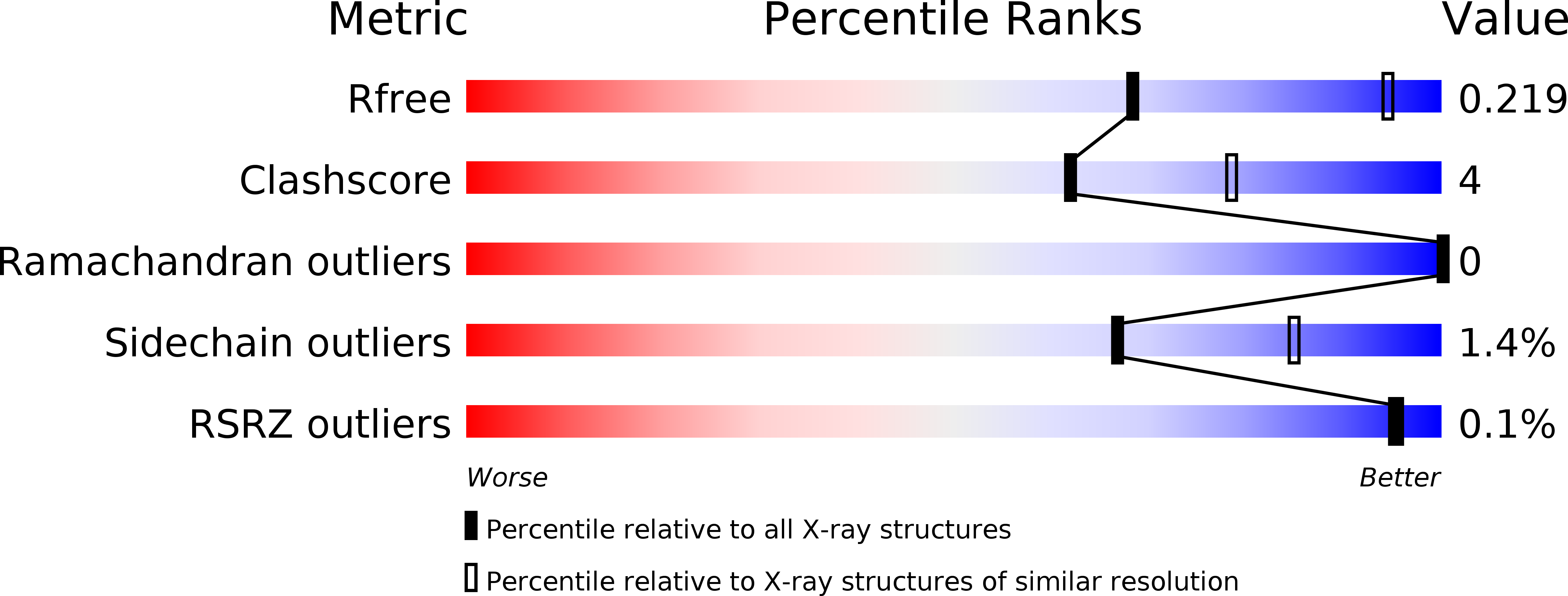
6DF2 image
Deposition Date 2018-05-13
Release Date 2018-11-28
Last Version Date 2024-10-30
Entry Detail
PDB ID:
6DF2
Keywords:
Title:
Improved anti-phosphotyrosine antibody 4G10-S5-4D5 Fab complexed with phosphotyrosine peptide
Biological Source:
Source Organism:
Host Organism:
Method Details:
Experimental Method:
Resolution:
2.60 Å
R-Value Free:
0.23
R-Value Work:
0.17
R-Value Observed:
0.17
Space Group:
C 1 2 1
Macromolecular Entities
Polymer Type:polypeptide(L)
Molecule:LEU-PTR-LEU
Chain IDs:C (auth: D), F (auth: C)
Chain Length:3
Number of Molecules:2
Biological Source:synthetic construct
Polymer Type:polypeptide(L)
Molecule:Anti-phosphotyrosine antibody c310-4D5 light chain
Chain IDs:B (auth: H), E (auth: B)
Chain Length:261
Number of Molecules:2
Biological Source:synthetic construct
Polymer Type:polypeptide(L)
Molecule:Anti-phosphotyrosine antibody c310-4D5 heavy chain
Chain IDs:A (auth: L), D (auth: A)
Chain Length:251
Number of Molecules:2
Biological Source:synthetic construct
Primary Citation
Engineering Improved Antiphosphotyrosine Antibodies Based on an Immunoconvergent Binding Motif.
J. Am. Chem. Soc. 140 16615 16624 (2018)
PMID: 30398859 DOI: 10.1021/jacs.8b08402

Abstact

Phosphotyrosine (pY) is one of the most highly studied posttranslational modifications that is responsible for tightly regulating many signaling pathways in eukaryotes. Pan-specific pY antibodies have emerged as powerful tools for understanding the role of these modifications. Nevertheless, structures have not been reported for pan-specific pY antibodies, greatly impeding the further development of tools for integrating this ubiquitous posttranslational modification using structure-guided designs. Here, we present the first crystal structures of two widely utilized pan-specific pY antibodies, PY20 and 4G10. The two antibodies, although developed independently from animal immunizations, have surprisingly similar modes of recognition of the phosphate group, implicating a generic binding structure among pan-specific pY antibodies. Sequence alignments revealed that many pY binding residues are predominant in the mouse V germline genes, which consequently led to the convergent antibodies. On the basis of the convergent structure, we designed a phage display library by lengthening the CDR-L3 loop with the aid of computational modeling. Panning with this library resulted in a series of 4G10 variants with 4 to 11-fold improvements in pY binding affinities. The crystal structure of one improved variant showed remarkable superposition to the computational model, where the lengthened CDR-L3 loop creates an additional hydrogen bond indirectly bound to the phosphate group via a water molecule. The engineered variants exhibited superior performance in Western blot and immunofluorescence.

Legend

Protein

Chemical

Disease

Primary Citation of related structures