Deposition Date
2018-05-11
Release Date
2018-12-19
Last Version Date
2023-10-11
Entry Detail
PDB ID:
6DEH
Keywords:
Title:
Structure of LpnE Effector Protein from Legionella pneumophila (sp. Philadelphia)
Biological Source:
Source Organism:
Legionella pneumophila subsp. pneumophila (Taxon ID: 272624)
Host Organism:
Method Details:
Experimental Method:
Resolution:
1.80 Å
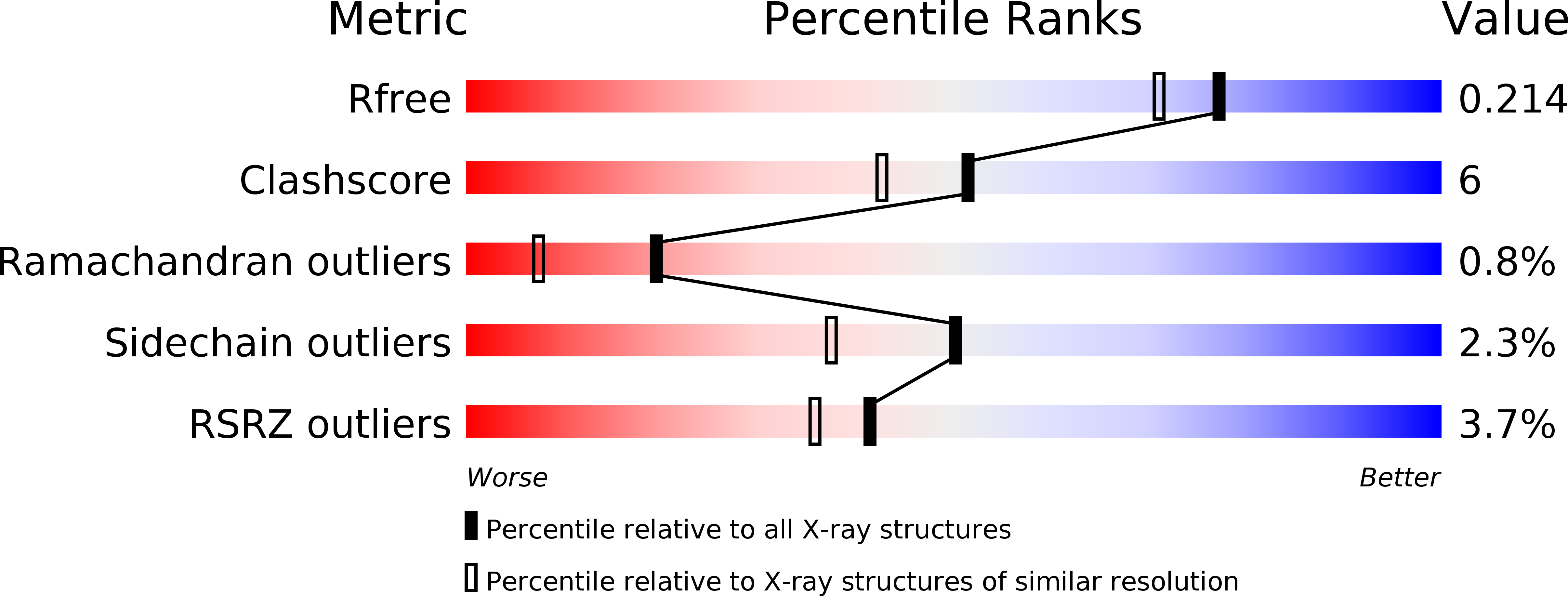
R-Value Free:
0.21
R-Value Work:
0.18
R-Value Observed:
0.18
Space Group:
P 21 21 21