6DAO image
Entry Detail
PDB ID:
6DAO
Keywords:
Title:
NahE WT selenomethionine
Biological Source:
Source Organism:
Host Organism:
PDB Version:
Deposition Date:
2018-05-01
Release Date:
2019-05-08
Method Details:
Experimental Method:
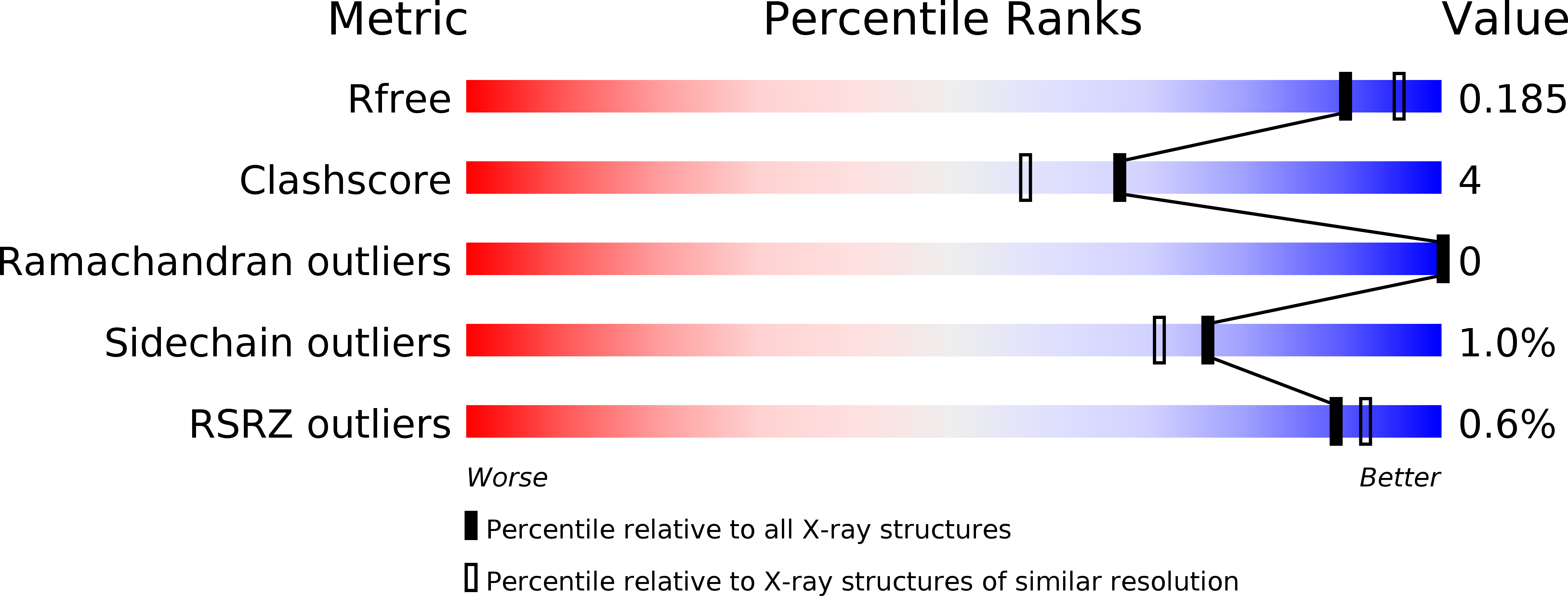
Resolution:
1.94 Å
R-Value Free:
0.18
R-Value Work:
0.14
R-Value Observed:
0.14
Space Group:
P 32 2 1
Macromolecular Entities
Polymer Type:polypeptide(L)
Description:Trans-O-hydroxybenzylidenepyruvate hydratase-aldolase
Chain IDs:A (auth: B), B (auth: A)
Chain Length:331
Number of Molecules:2
Biological Source:Pseudomonas putida
Modified Residue
Compound ID Chain ID Parent Comp ID Details 2D Image
MSE A MET modified residue
Primary Citation
Structural Characterization of the Hydratase-Aldolases, NahE and PhdJ: Implications for the Specificity, Catalysis, and N-Acetylneuraminate Lyase Subgroup of the Aldolase Superfamily.
Biochemistry 57 3524 3536 (2018)
PMID: 29856600 DOI: 10.1021/acs.biochem.8b00532

Abstact

NahE and PhdJ are bifunctional hydratase-aldolases in bacterial catabolic pathways for naphthalene and phenanthrene, respectively. Bacterial species with these pathways can use polycyclic aromatic hydrocarbons (PAHs) as sole sources of carbon and energy. Because of the harmful properties of PAHs and their widespread distribution and persistence in the environment, there is great interest in understanding these degradative pathways, including the mechanisms and specificities of the enzymes found in the pathways. This knowledge can be used to develop and optimize bioremediation techniques. Although hydratase-aldolases catalyze a major step in the PAH degradative pathways, their mechanisms are poorly understood. Sequence analysis identified NahE and PhdJ as members of the N-acetylneuraminate lyase (NAL) subgroup in the aldolase superfamily. Both have a conserved lysine and tyrosine (for Schiff base formation) as well as a GXXGE motif (to bind the pyruvoyl carboxylate group). Herein, we report the structures of NahE, PhdJ, and PhdJ covalently bound to substrate via a Schiff base. Structural analysis and dynamic light scattering experiments show that both enzymes are tetramers. A hydrophobic helix insert, present in the active sites of NahE and PhdJ, might differentiate them from other NAL subgroup members. The individual specificities of NahE and PhdJ are governed by Asn-281/Glu-285 and Ser-278/Asp-282, respectively. Finally, the PhdJ complex structure suggests a potential mechanism for hydration of substrate and subsequent retro-aldol fission. The combined findings fill a gap in our mechanistic understanding of these enzymes and their place in the NAL subgroup.

Legend

Protein

Chemical

Disease

Primary Citation of related structures