6DAM image
Deposition Date 2018-05-01
Release Date 2018-09-05
Last Version Date 2024-11-13
Entry Detail
PDB ID:
6DAM
Keywords:
Title:
Crystal structure of lanthanide-dependent methanol dehydrogenase XoxF from Methylomicrobium buryatense 5G
Biological Source:
Source Organism:
Method Details:
Experimental Method:
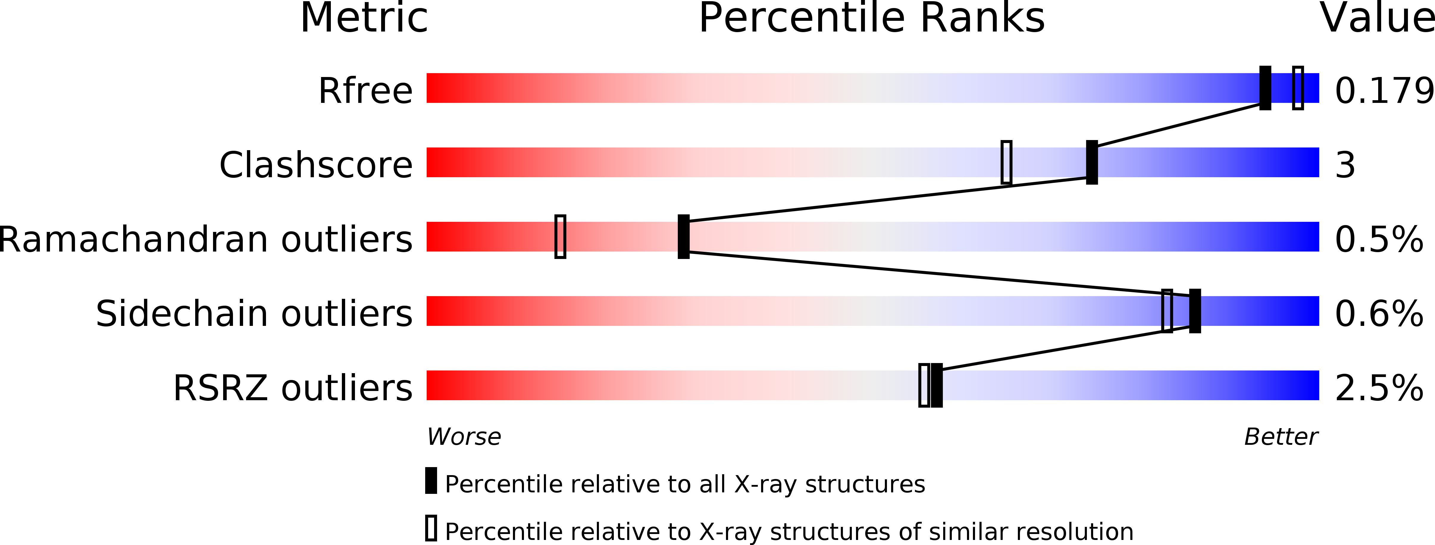
Resolution:
1.85 Å
R-Value Free:
0.18
R-Value Work:
0.15
R-Value Observed:
0.15
Space Group:
C 2 2 21
Macromolecular Entities
Structures with similar UniProt ID
Protein Blast
Polymer Type:polypeptide(L)
Molecule:Lanthanide-dependent methanol dehydrogenase XoxF
Chain IDs:A
Chain Length:617
Number of Molecules:1
Biological Source:Methylomicrobium buryatense 5G
Primary Citation
Structure and function of the lanthanide-dependent methanol dehydrogenase XoxF from the methanotroph Methylomicrobium buryatense 5GB1C.
J. Biol. Inorg. Chem. 23 1037 1047 (2018)
PMID: 30132076 DOI: 10.1007/s00775-018-1604-2

Abstact

In methylotrophic bacteria, which use one-carbon (C1) compounds as a carbon source, methanol is oxidized by pyrroloquinoline quinone (PQQ)-dependent methanol dehydrogenase (MDH) enzymes. Methylotrophic genomes generally encode two distinct MDHs, MxaF and XoxF. MxaF is a well-studied, calcium-dependent heterotetrameric enzyme whereas XoxF is a lanthanide-dependent homodimer. Recent studies suggest that XoxFs are likely the functional MDHs in many environments. In methanotrophs, methylotrophs that utilize methane, interactions between particulate methane monooxygenase (pMMO) and MxaF have been detected. To investigate the possibility of interactions between pMMO and XoxF, XoxF was isolated from the methanotroph Methylomicrobium buryatense 5GB1C (5G-XoxF). Purified 5G-XoxF exhibits a specific activity of 0.16 μmol DCPIP reduced min-1 mg-1. The 1.85 Å resolution crystal structure reveals a La(III) ion in the active site, in contrast to the calcium ion in MxaF. The overall fold is similar to other MDH structures, but 5G-XoxF is a monomer in solution. An interaction between 5G-XoxF and its cognate pMMO was detected by biolayer interferometry, with a KD value of 50 ± 17 μM. These results suggest an alternative model of MDH-pMMO association, in which a XoxF monomer may bind to pMMO, and underscore the potential importance of lanthanide-dependent MDHs in biological methane oxidation.

Legend

Protein

Chemical

Disease

Primary Citation of related structures