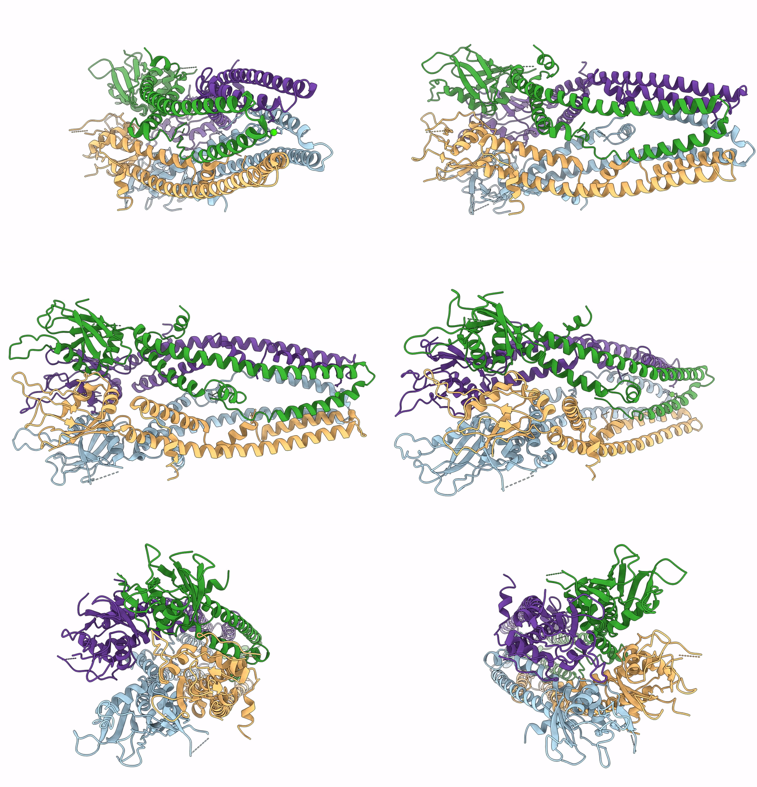
6D7W image
Deposition Date 2018-04-25
Release Date 2018-07-11
Last Version Date 2025-06-04
Entry Detail
PDB ID:
6D7W
Title:
Cryo-EM structure of the mitochondrial calcium uniporter from N. fischeri at 3.8 Angstrom resolution
Biological Source:
Source Organism:
Method Details:
Experimental Method:
Resolution:
3.80 Å
Aggregation State:
PARTICLE
Reconstruction Method:
SINGLE PARTICLE
Macromolecular Entities
Structures with similar UniProt ID
Protein Blast
Polymer Type:polypeptide(L)
Molecule:Mitochondrial calcium uniporter
Gene (Uniprot):mcu
Chain IDs:A, B (auth: C), C (auth: B), D
Chain Length:416
Number of Molecules:4
Biological Source:Aspergillus fischeri
Ligand Molecules
Primary Citation
Cryo-EM structure of a fungal mitochondrial calcium uniporter.
Nature 559 570 574 (2018)
PMID: 29995855 DOI: 10.1038/s41586-018-0333-6

Abstact

The mitochondrial calcium uniporter (MCU) is a highly selective calcium channel localized to the inner mitochondrial membrane. Here, we describe the structure of an MCU orthologue from the fungus Neosartorya fischeri (NfMCU) determined to 3.8 Å resolution by phase-plate cryo-electron microscopy. The channel is a homotetramer with two-fold symmetry in its amino-terminal domain (NTD) that adopts a similar structure to that of human MCU. The NTD assembles as a dimer of dimers to form a tetrameric ring that connects to the transmembrane domain through an elongated coiled-coil domain. The ion-conducting pore domain maintains four-fold symmetry, with the selectivity filter positioned at the start of the pore-forming TM2 helix. The aspartate and glutamate sidechains of the conserved DIME motif are oriented towards the central axis and separated by one helical turn. The structure of NfMCU offers insights into channel assembly, selective calcium permeation, and inhibitor binding.

Legend

Protein

Chemical

Disease

Primary Citation of related structures