6D7K image
Entry Detail
PDB ID:
6D7K
Title:
Complex structure of Methane monooxygenase hydroxylase in complex with inhibitory subunit
Biological Source:
Source Organism:
Host Organism:
PDB Version:
Deposition Date:
2018-04-24
Release Date:
2019-06-26
Method Details:
Experimental Method:
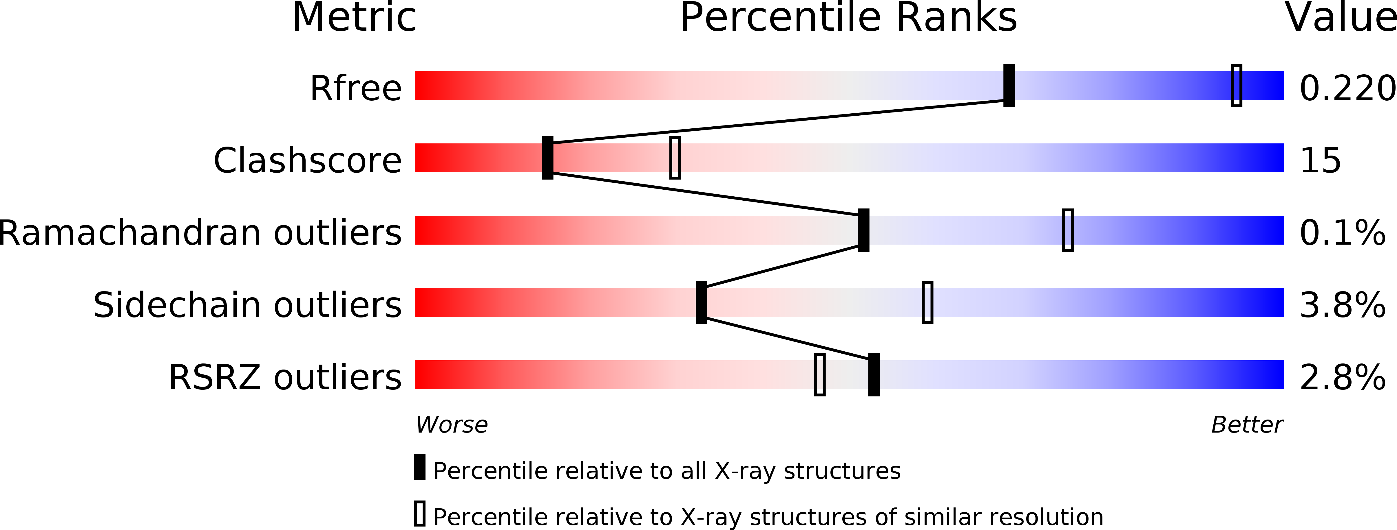
Resolution:
2.60 Å
R-Value Free:
0.22
R-Value Work:
0.17
R-Value Observed:
0.17
Space Group:
C 1 2 1
Macromolecular Entities
Polymer Type:polypeptide(L)
Description:Methane monooxygenase hydroxylase, MmoX1
Chain IDs:A, E
Chain Length:526
Number of Molecules:2
Biological Source:Methylosinus sporium
Polymer Type:polypeptide(L)
Description:Methane monooxygenase hydroxylase, MmoY
Chain IDs:B, F
Chain Length:395
Number of Molecules:2
Biological Source:Methylosinus sporium
Polymer Type:polypeptide(L)
Description:Methane monooxygenase hydroxylase, MmoZ
Chain IDs:C, G
Chain Length:169
Number of Molecules:2
Biological Source:Methylosinus sporium
Polymer Type:polypeptide(L)
Description:Methane monooxygenase hydroxylase, MmoD
Chain IDs:D, H
Chain Length:114
Number of Molecules:2
Biological Source:Methylosinus sporium
Primary Citation
MMOD-induced structural changes of hydroxylase in soluble methane monooxygenase.
Sci Adv 5 eaax0059 eaax0059 (2019)
PMID: 31616787 DOI: 10.1126/sciadv.aax0059

Abstact

Soluble methane monooxygenase in methanotrophs converts methane to methanol under ambient conditions. The maximum catalytic activity of hydroxylase (MMOH) is achieved through the interplay of its regulatory protein (MMOB) and reductase. An additional auxiliary protein, MMOD, functions as an inhibitor of MMOH; however, its inhibitory mechanism remains unknown. Here, we report the crystal structure of the MMOH-MMOD complex from Methylosinus sporium strain 5 (2.6 Å). Its structure illustrates that MMOD associates with the canyon region of MMOH where MMOB binds. Although MMOD and MMOB recognize the same binding site, each binding component triggers different conformational changes toward MMOH, which then respectively lead to the inhibition and activation of MMOH. Particularly, MMOD binding perturbs the di-iron geometry by inducing two major MMOH conformational changes, i.e., MMOH β subunit disorganization and subsequent His147 dissociation with Fe1 coordination. Furthermore, 1,6-hexanediol, a mimic of the products of sMMO, reveals the substrate access route.

Legend

Protein

Chemical

Disease

Primary Citation of related structures