6D6B image
Entry Detail
PDB ID:
6D6B
Title:
The structure of ligand binding domain of LasR in complex with TP-1 homolog, compound 11
Biological Source:
PDB Version:
Deposition Date:
2018-04-20
Release Date:
2018-08-08
Method Details:
Experimental Method:
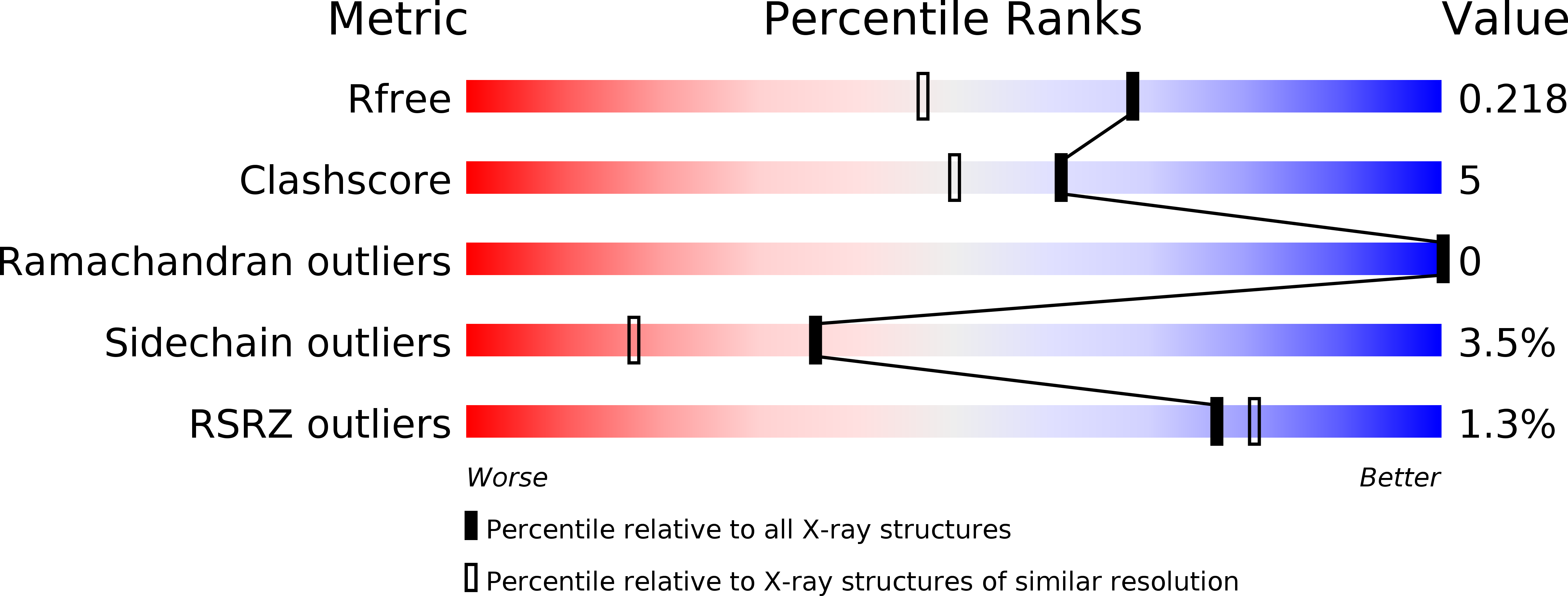
Resolution:
1.70 Å
R-Value Free:
0.20
R-Value Work:
0.17
R-Value Observed:
0.18
Space Group:
P 1 21 1
Macromolecular Entities
Polymer Type:polypeptide(L)
Description:Transcriptional activator protein LasR
Chain IDs:A, B, C, D, E, F, G, H
Chain Length:170
Number of Molecules:8
Biological Source:Pseudomonas aeruginosa (strain ATCC 15692 / DSM 22644 / CIP 104116 / JCM 14847 / LMG 12228 / 1C / PRS 101 / PAO1)
Ligand Molecules
Primary Citation
Structural and Biochemical Studies of Non-native Agonists of the LasR Quorum-Sensing Receptor Reveal an L3 Loop "Out" Conformation for LasR.
Cell Chem Biol 25 1128 1139.e3 (2018)
PMID: 30033130 DOI: 10.1016/j.chembiol.2018.06.007

Abstact

Chemical strategies to block quorum sensing (QS) could provide a route to attenuate virulence in bacterial pathogens. Considerable research has focused on this approach in Pseudomonas aeruginosa, which uses the LuxR-type receptor LasR to regulate much of its QS network. Non-native ligands that antagonize LasR have been developed, yet we have little understanding of the mode by which these compounds interact with LasR and alter its function, as the receptor is unstable in their presence. Herein, we report an approach to circumvent this challenge through the study of a series of synthetic LasR agonists with varying levels of potency. Structural investigations of these ligands with the LasR ligand-binding domain reveal that certain agonists can enforce a conformation that deviates from that observed for other, often more potent agonists. These results, when combined with cell-based and biophysical analyses, suggest a functional model for LasR that could guide future ligand design.

Legend

Protein

Chemical

Disease

Primary Citation of related structures