Deposition Date
2018-04-18
Release Date
2018-10-17
Last Version Date
2023-10-04
Entry Detail
PDB ID:
6D4F
Keywords:
Title:
Crystal structure of PTP epsilon D2 domain (A455N/V457Y/E597D)
Biological Source:
Source Organism(s):
Homo sapiens (Taxon ID: 9606)
Expression System(s):
Method Details:
Experimental Method:
Resolution:
1.91 Å
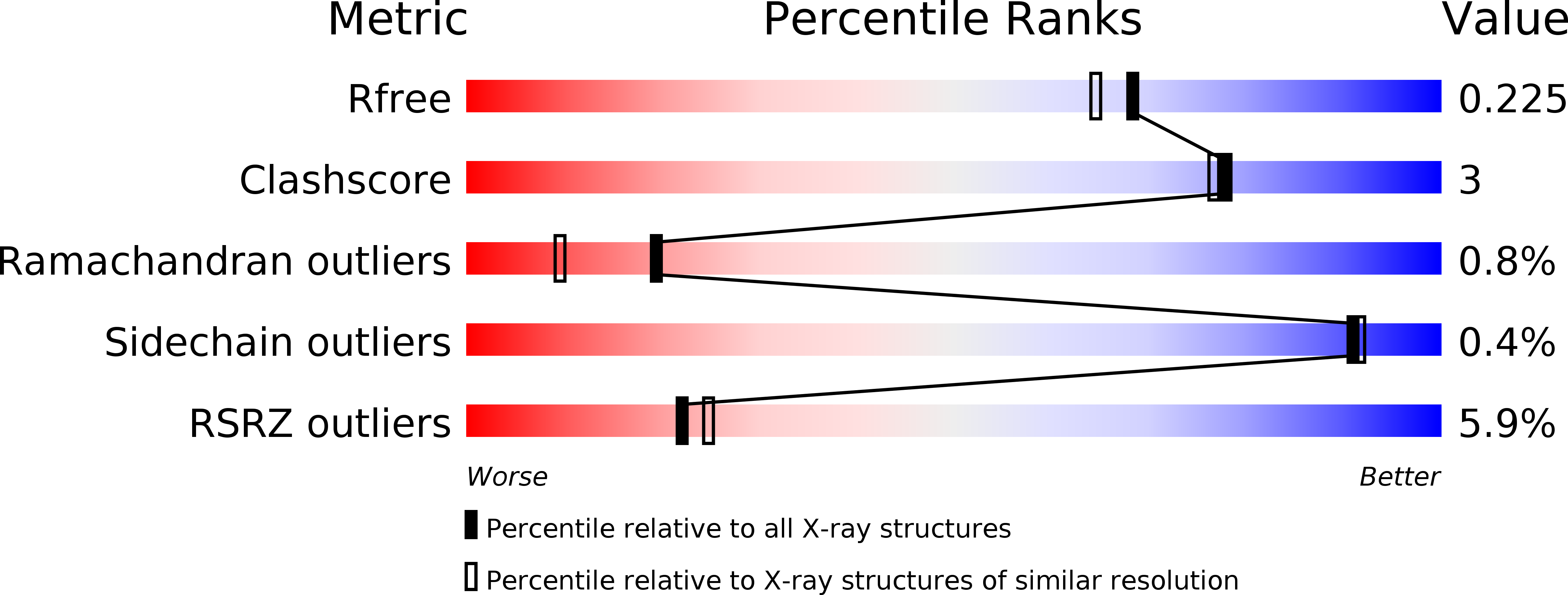
R-Value Free:
0.22
R-Value Work:
0.19
R-Value Observed:
0.19
Space Group:
P 21 21 21