6D42 image
Deposition Date 2018-04-17
Release Date 2018-07-11
Last Version Date 2024-11-20
Entry Detail
PDB ID:
6D42
Title:
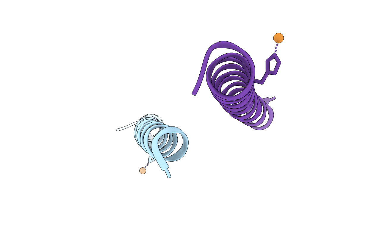
Crystal structure of the KCa3.1 C-terminal four-helix bundle (with copper)
Biological Source:
Source Organism:
Homo sapiens (Taxon ID: 9606)
Host Organism:
Method Details:
Experimental Method:
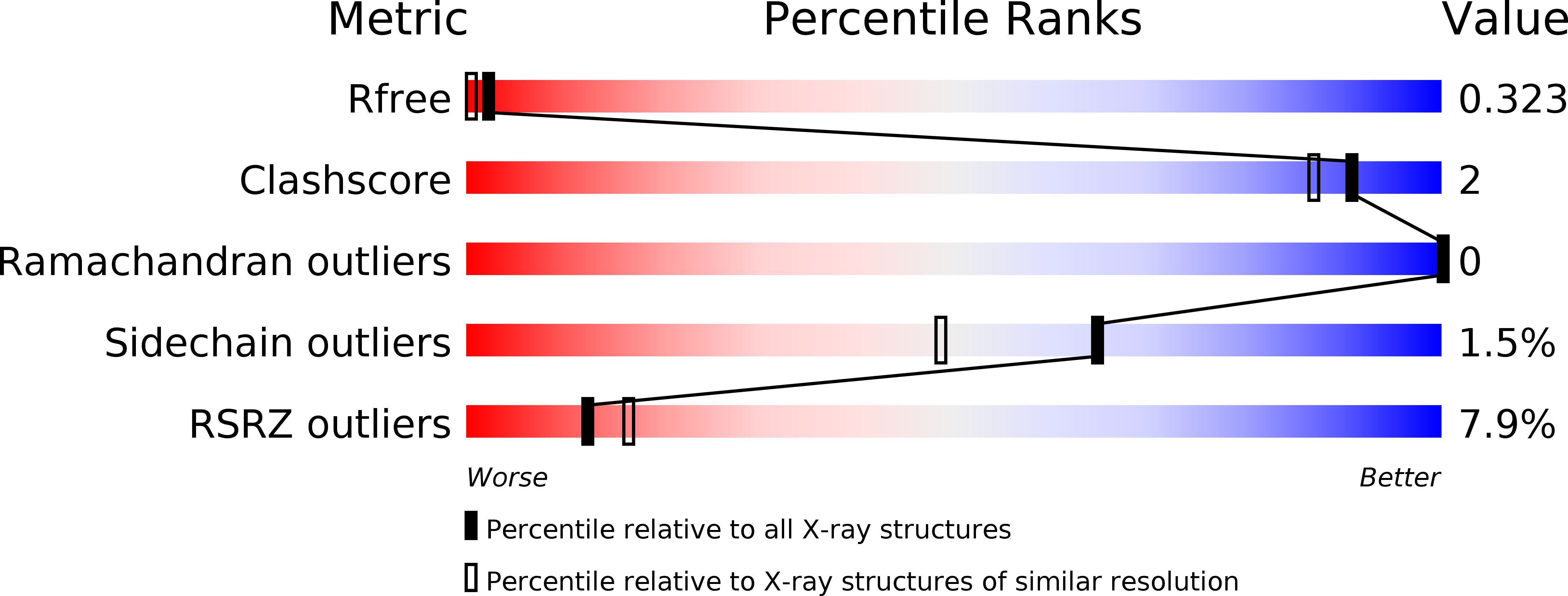
Resolution:
1.75 Å
R-Value Free:
0.32
R-Value Work:
0.25
R-Value Observed:
0.25
Space Group:
P 4 21 2
Macromolecular Entities
Polymer Type:polypeptide(L)
Molecule:Intermediate conductance calcium-activated potassium channel protein 4
Gene (Uniprot):KCNN4
Chain IDs:A, B
Chain Length:42
Number of Molecules:2
Biological Source:Homo sapiens
Modified Residue
Compound ID Chain ID Parent Comp ID Details 2D Image
MSE A MET modified residue
Ligand Molecules
Primary Citation
Crystal structure of the C-terminal four-helix bundle of the potassium channel KCa3.1.
PLoS ONE 13 e0199942 e0199942 (2018)
PMID: 29953543 DOI: 10.1371/journal.pone.0199942

Abstact

KCa3.1 (also known as SK4 or IK1) is a mammalian intermediate-conductance potassium channel that plays a critical role in the activation of T cells, B cells, and mast cells, effluxing potassium ions to maintain a negative membrane potential for influxing calcium ions. KCa3.1 shares primary sequence similarity with three other (low-conductance) potassium channels: KCa2.1, KCa2.2, and KCa2.3 (also known as SK1-3). These four homotetrameric channels bind calmodulin (CaM) in the cytoplasmic region, and calcium binding to CaM triggers channel activation. Unique to KCa3.1, activation also requires phosphorylation of a single histidine residue, His358, in the cytoplasmic region, which relieves copper-mediated inhibition of the channel. Near the cytoplasmic C-terminus of KCa3.1 (and KCa2.1-2.3), secondary-structure analysis predicts the presence of a coiled-coil/heptad repeat. Here, we report the crystal structure of the C-terminal coiled-coil region of KCa3.1, which forms a parallel four-helix bundle, consistent with the tetrameric nature of the channel. Interestingly, the four copies of a histidine residue, His389, in an 'a' position within the heptad repeat, are observed to bind a copper ion along the four-fold axis of the bundle. These results suggest that His358, the inhibitory histidine in KCa3.1, might coordinate a copper ion through a similar binding mode.

Legend

Protein

Chemical

Disease

Primary Citation of related structures