6CWH image
Entry Detail
PDB ID:
6CWH
Title:
Crystal structure of SpaA-SLH in complex with 4,6-Pyr-beta-D-ManNAcOMe (P1)
Biological Source:
Source Organism:
PDB Version:
Deposition Date:
2018-03-30
Release Date:
2018-08-15
Method Details:
Experimental Method:
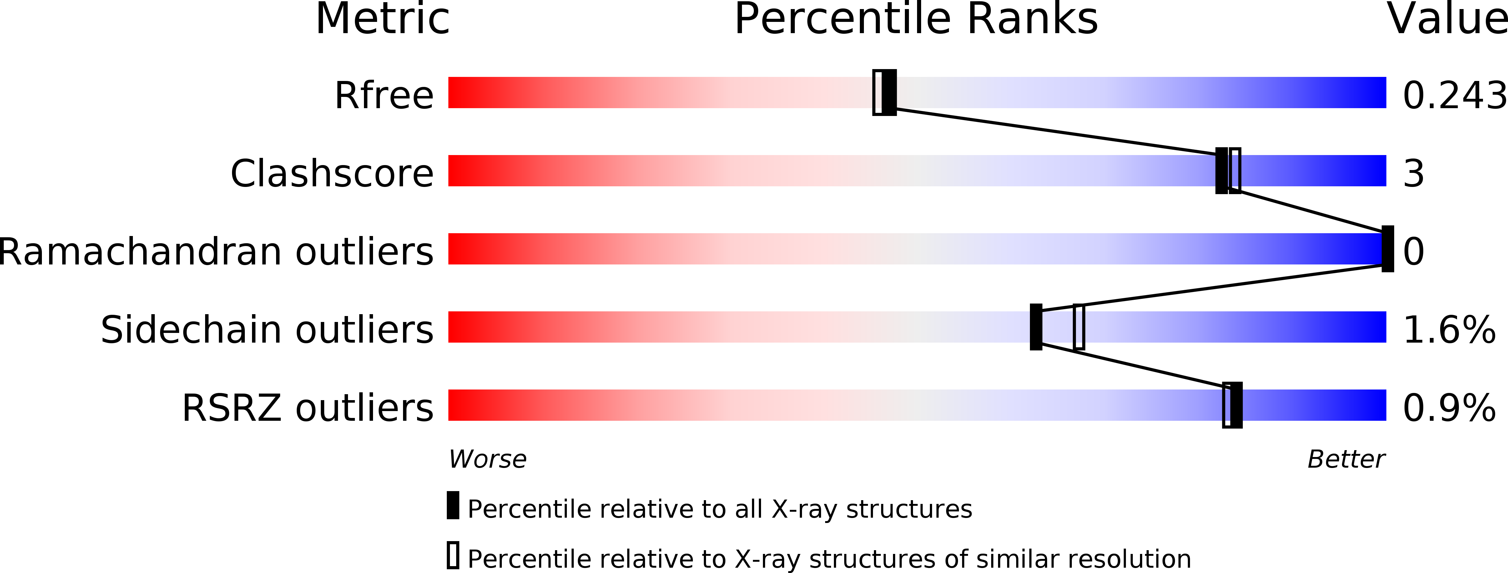
Resolution:
2.00 Å
R-Value Free:
0.23
R-Value Work:
0.18
R-Value Observed:
0.18
Space Group:
P 1
Macromolecular Entities
Polymer Type:polypeptide(L)
Description:Surface (S-) layer glycoprotein
Chain IDs:A, B, C, D
Chain Length:182
Number of Molecules:4
Biological Source:Paenibacillus alvei
Ligand Molecules
Primary Citation
Structural basis of cell wall anchoring by SLH domains in Paenibacillus alvei.
Nat Commun 9 3120 3120 (2018)
PMID: 30087354 DOI: 10.1038/s41467-018-05471-3

Abstact

Self-assembling protein surface (S-) layers are common cell envelope structures of prokaryotes and have critical roles from structural maintenance to virulence. S-layers of Gram-positive bacteria are often attached through the interaction of S-layer homology (SLH) domain trimers with peptidoglycan-linked secondary cell wall polymers (SCWPs). Here we present an in-depth characterization of this interaction, with co-crystal structures of the three consecutive SLH domains from the Paenibacillus alvei S-layer protein SpaA with defined SCWP ligands. The most highly conserved SLH domain residue SLH-Gly29 is shown to enable a peptide backbone flip essential for SCWP binding in both biophysical and cellular experiments. Furthermore, we find that a significant domain movement mediates binding by two different sites in the SLH domain trimer, which may allow anchoring readjustment to relieve S-layer strain caused by cell growth and division.

Legend

Protein

Chemical

Disease

Primary Citation of related structures