6CPB image
Deposition Date 2018-03-13
Release Date 2018-05-16
Last Version Date 2023-10-04
Entry Detail
PDB ID:
6CPB
Keywords:
Title:
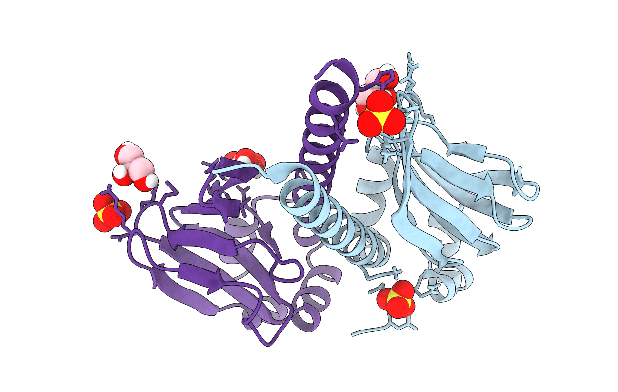
Crystal structure of the heme domain of CooA from Carboxydothermus hydrogenoformans
Biological Source:
Source Organism:
Host Organism:
Method Details:
Experimental Method:
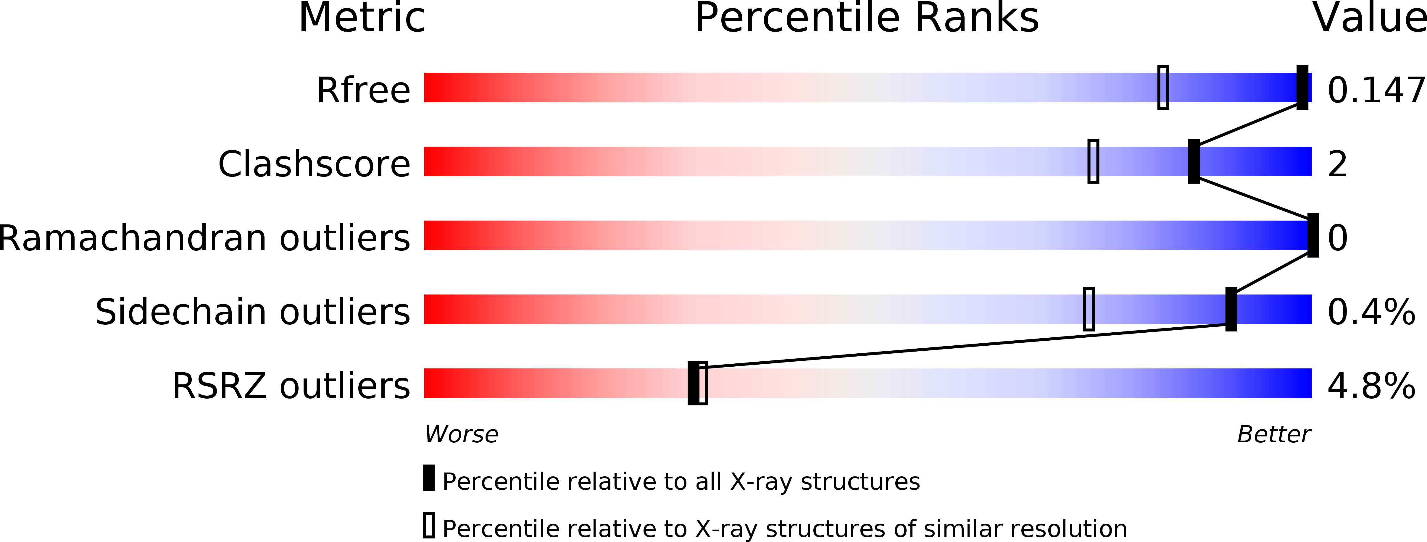
Resolution:
1.16 Å
R-Value Free:
0.14
R-Value Work:
0.12
R-Value Observed:
0.12
Space Group:
P 21 21 21
Macromolecular Entities
Structures with similar UniProt ID
Protein Blast
Polymer Type:polypeptide(L)
Molecule:Carbon monoxide oxidation system transcription regulator CooA-1
Gene (Uniprot):cooA-1
Chain IDs:A, B
Chain Length:141
Number of Molecules:2
Biological Source:Carboxydothermus hydrogenoformans
Primary Citation
Testing the N-Terminal Velcro Model of CooA Carbon Monoxide Activation.
Biochemistry 57 3059 3064 (2018)
PMID: 29708736 DOI: 10.1021/acs.biochem.8b00359

Abstact

CooAs are dimeric bacterial CO-sensing transcription factors that activate a series of enzymes responsible for CO oxidation. The crystal structure of Rhodospirillum rubrum (rrCooA) shows that the N-terminal Pro from monomer A of the dimer coordinates the heme of monomer B that locks rrCooA in the "off" state. When CO binds, it is postulated that the Pro is replaced with CO, resulting in a very large reorientation of the DNA binding domains required for specific binding to DNA. Crystal structures of the closely related CooA from Carboxydothermus hydrogenoformans (chCooA) are available, and in one of these, the CO-bound on-state indicates that the N-terminal region that is displaced when CO binds provides contacts between the heme and DNA binding domains that hold the DNA binding domain in position for DNA binding. This has been termed the N-terminal velcro model of CooA activation. The study presented here tests this hypothesis by generating a disulfide mutant that covalently locks chCooA in the on-state. A simple fluorescence assay was used to measure DNA binding, and the S-S mutant was found to be in the on-state even without CO. We also determined the high-resolution crystal structure of the apo-heme domain, and the resulting structure is very similar to the holo-heme-bound structure. This result shows that the heme binding motif forms a stable structure without heme or the DNA binding domain.

Legend

Protein

Chemical

Disease

Primary Citation of related structures