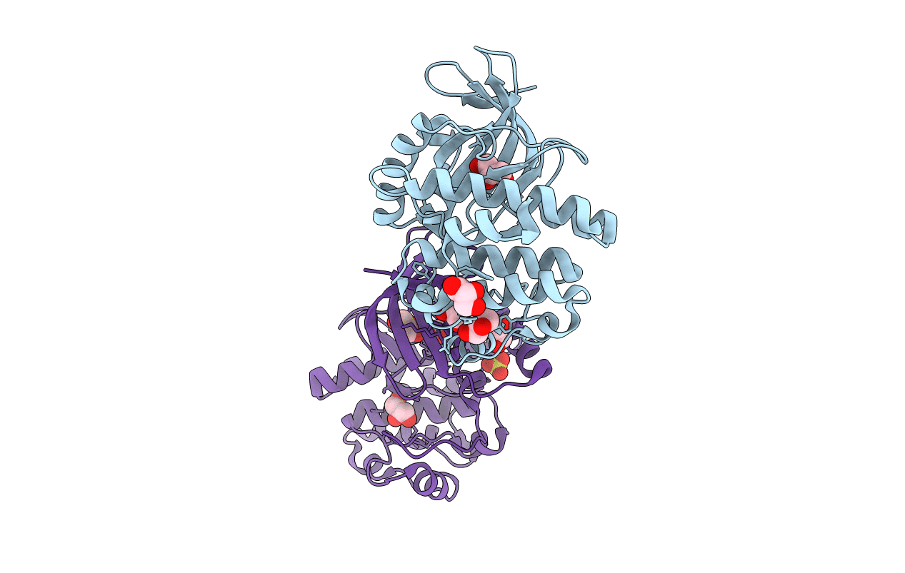
6CN9 image
Entry Detail
PDB ID:
6CN9
Keywords:
Title:
Crystal structure of the Kinase domain of WNK1
Biological Source:
Source Organism:
Host Organism:
PDB Version:
Deposition Date:
2018-03-07
Release Date:
2019-03-27
Method Details:
Experimental Method:
Resolution:
1.80 Å
R-Value Free:
0.21
R-Value Work:
0.16
R-Value Observed:
0.16
Space Group:
P 1
Macromolecular Entities
Polymer Type:polypeptide(L)
Description:Serine/threonine-protein kinase WNK1
Chain IDs:A, B
Chain Length:290
Number of Molecules:2
Biological Source:Rattus norvegicus
Modified Residue
Compound ID Chain ID Parent Comp ID Details 2D Image
MSE A MET modified residue
Primary Citation
Crystal structure of the kinase domain of WNK1, a kinase that causes a hereditary form of hypertension.
Structure 12 1303 1311 (2004)
PMID: 15242606 DOI: 10.1016/j.str.2004.04.014

Abstact

WNK kinases comprise a small group of unique serine/threonine protein kinases that have been genetically linked to pseudohypoaldosteronism type II, an autosomal dominant form of hypertension. Here we present the structure of the kinase domain of WNK1 at 1.8 A resolution, solved in a low activity conformation. A lysine residue (Lys-233) is found in the active site emanating from strand beta2 rather than strand beta3 as in other protein kinases. The activation loop adopts a unique well-folded inactive conformation. The conformations of the P+1 specificity pocket, the placement of the conserved active site threonine (Thr-386), and the exterior placement of helix C, contribute to the low activity state. By homology modeling, we identified two hydrophobic residues in the substrate-binding groove that contribute to substrate specificity. The structure of the WNK1 catalytic domain, with its unique active site, may help in the design of therapeutic reagents for the treatment of hypertension.

Legend

Protein

Chemical

Disease

Primary Citation of related structures