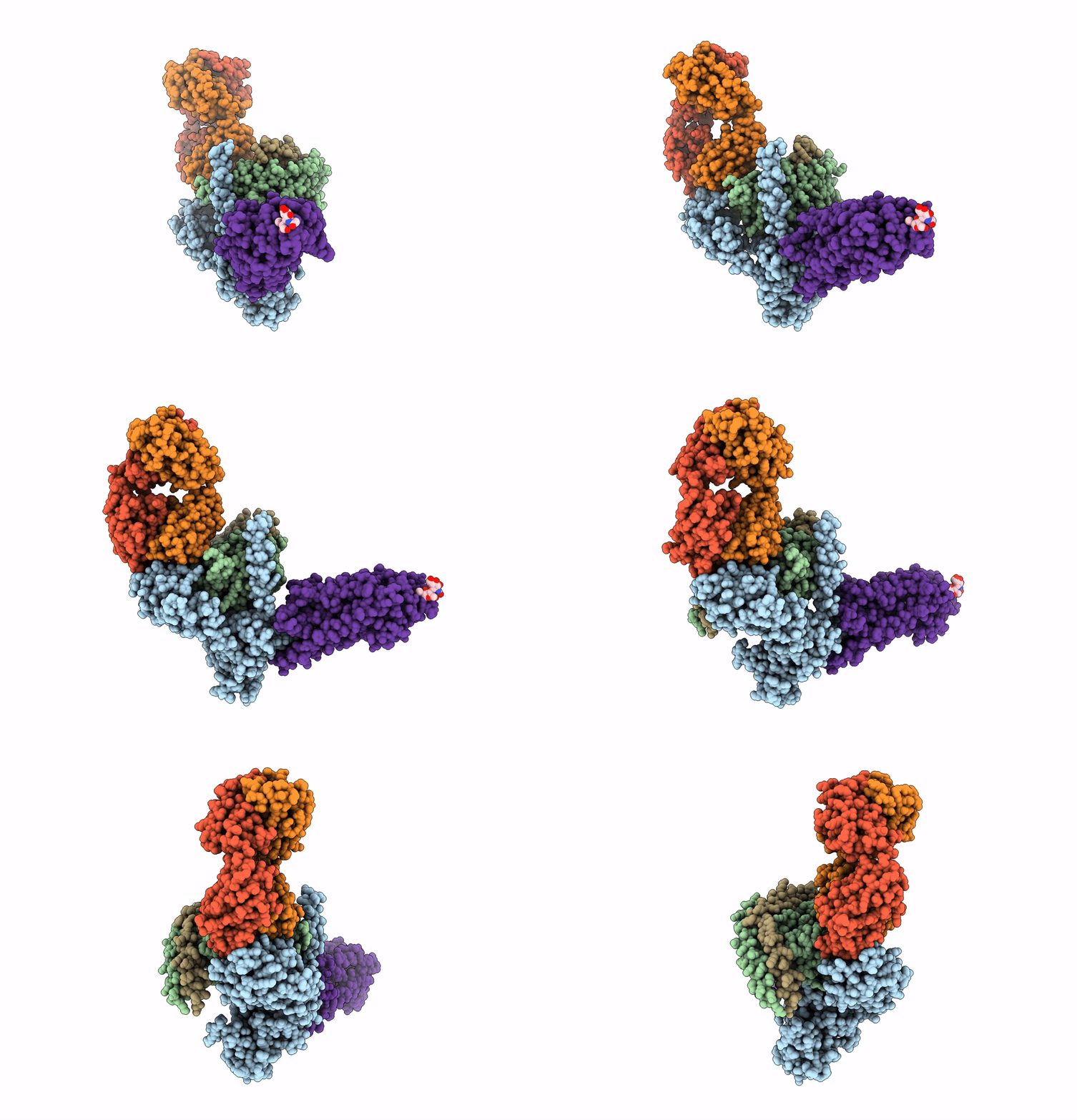
6CMO image
Entry Detail
PDB ID:
6CMO
EMDB ID:
Title:
Rhodopsin-Gi complex
Biological Source:
PDB Version:
Deposition Date:
2018-03-05
Release Date:
2018-06-20
Method Details:
Experimental Method:
Resolution:
4.50 Å
Aggregation State:
PARTICLE
Reconstruction Method:
SINGLE PARTICLE
Macromolecular Entities
Polymer Type:polypeptide(L)
Description:Guanine nucleotide-binding protein G(i) subunit alpha-1
Chain IDs:B (auth: A)
Chain Length:354
Number of Molecules:1
Biological Source:Homo sapiens
Polymer Type:polypeptide(L)
Description:Guanine nucleotide-binding protein G(I)/G(S)/G(T) subunit beta-1
Chain IDs:C (auth: B)
Chain Length:345
Number of Molecules:1
Biological Source:Rattus norvegicus
Polymer Type:polypeptide(L)
Description:Guanine nucleotide-binding protein G(I)/G(S)/G(O) subunit gamma-2
Chain IDs:D (auth: G)
Chain Length:68
Number of Molecules:1
Biological Source:Bos taurus
Polymer Type:polypeptide(L)
Description:Fab Heavy chain
Chain IDs:F (auth: H)
Chain Length:239
Number of Molecules:1
Biological Source:synthetic construct
Polymer Type:polypeptide(L)
Description:Fab light chain
Chain IDs:E (auth: L)
Chain Length:215
Number of Molecules:1
Biological Source:synthetic construct
Polymer Type:polypeptide(L)
Description:chimera protein of Soluble cytochrome b562 and Rhodopsin
Chain IDs:A (auth: R)
Chain Length:463
Number of Molecules:1
Biological Source:Escherichia coli, Homo sapiens
Ligand Molecules
Primary Citation
Cryo-EM structure of human rhodopsin bound to an inhibitory G protein.
Nature 558 553 558 (2018)
PMID: 29899450 DOI: 10.1038/s41586-018-0215-y

Abstact

G-protein-coupled receptors comprise the largest family of mammalian transmembrane receptors. They mediate numerous cellular pathways by coupling with downstream signalling transducers, including the hetrotrimeric G proteins Gs (stimulatory) and Gi (inhibitory) and several arrestin proteins. The structural mechanisms that define how G-protein-coupled receptors selectively couple to a specific type of G protein or arrestin remain unknown. Here, using cryo-electron microscopy, we show that the major interactions between activated rhodopsin and Gi are mediated by the C-terminal helix of the Gi α-subunit, which is wedged into the cytoplasmic cavity of the transmembrane helix bundle and directly contacts the amino terminus of helix 8 of rhodopsin. Structural comparisons of inactive, Gi-bound and arrestin-bound forms of rhodopsin with inactive and Gs-bound forms of the β2-adrenergic receptor provide a foundation to understand the unique structural signatures that are associated with the recognition of Gs, Gi and arrestin by activated G-protein-coupled receptors.

Legend

Protein

Chemical

Disease

Primary Citation of related structures