Deposition Date
2018-03-05
Release Date
2018-06-06
Last Version Date
2023-10-04
Entry Detail
PDB ID:
6CMN
Keywords:
Title:
Co-Crystal Structure of HIV-1 TAR Bound to Lab-Evolved RRM TBP6.7
Biological Source:
Source Organism(s):
Oryctolagus cuniculus (Taxon ID: 9986)
Human immunodeficiency virus 1 (Taxon ID: 11676)
Human immunodeficiency virus 1 (Taxon ID: 11676)
Expression System(s):
Method Details:
Experimental Method:
Resolution:
1.80 Å
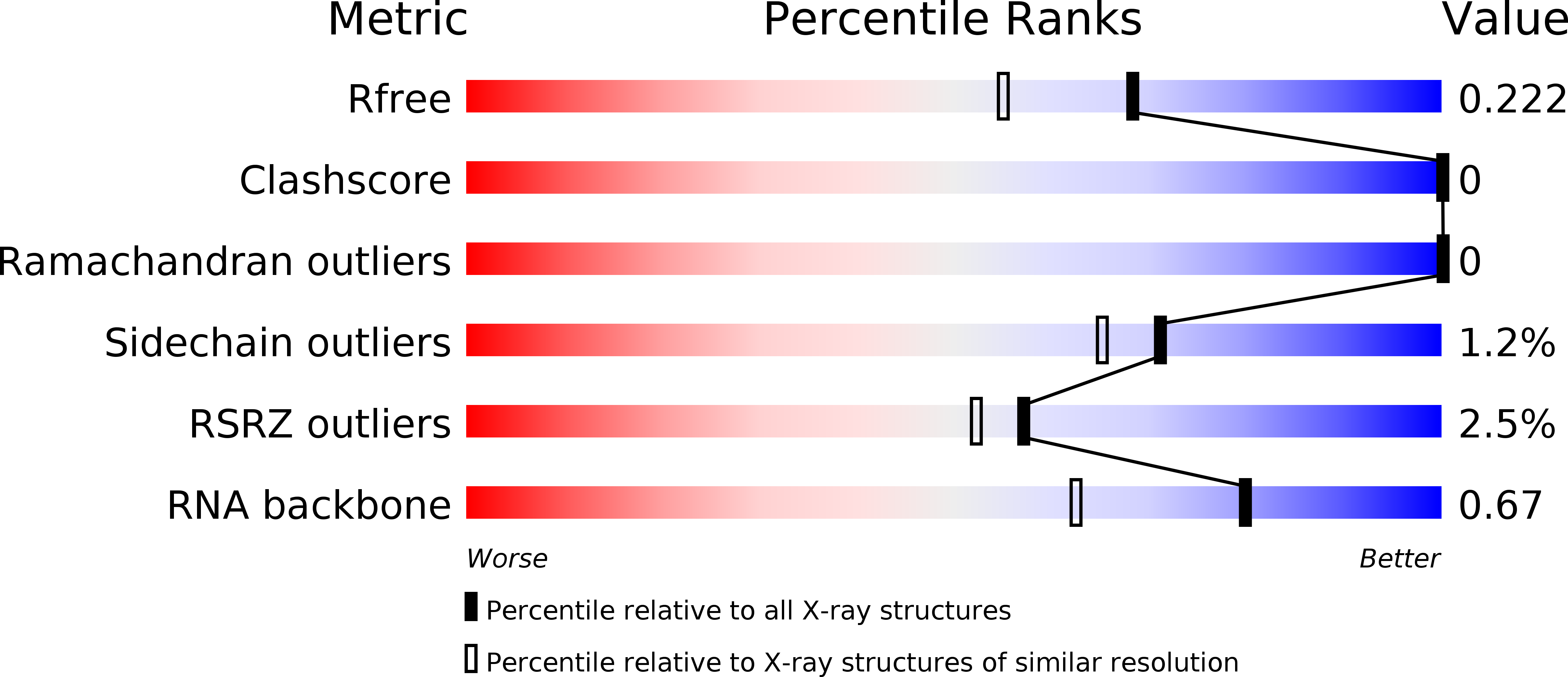
R-Value Free:
0.22
R-Value Work:
0.18
R-Value Observed:
0.19
Space Group:
P 43 21 2