6CMK image
Entry Detail
PDB ID:
6CMK
Title:
Crystal structure of Citrobacter koseri AztD
Biological Source:
PDB Version:
Deposition Date:
2018-03-05
Release Date:
2019-03-13
Method Details:
Experimental Method:
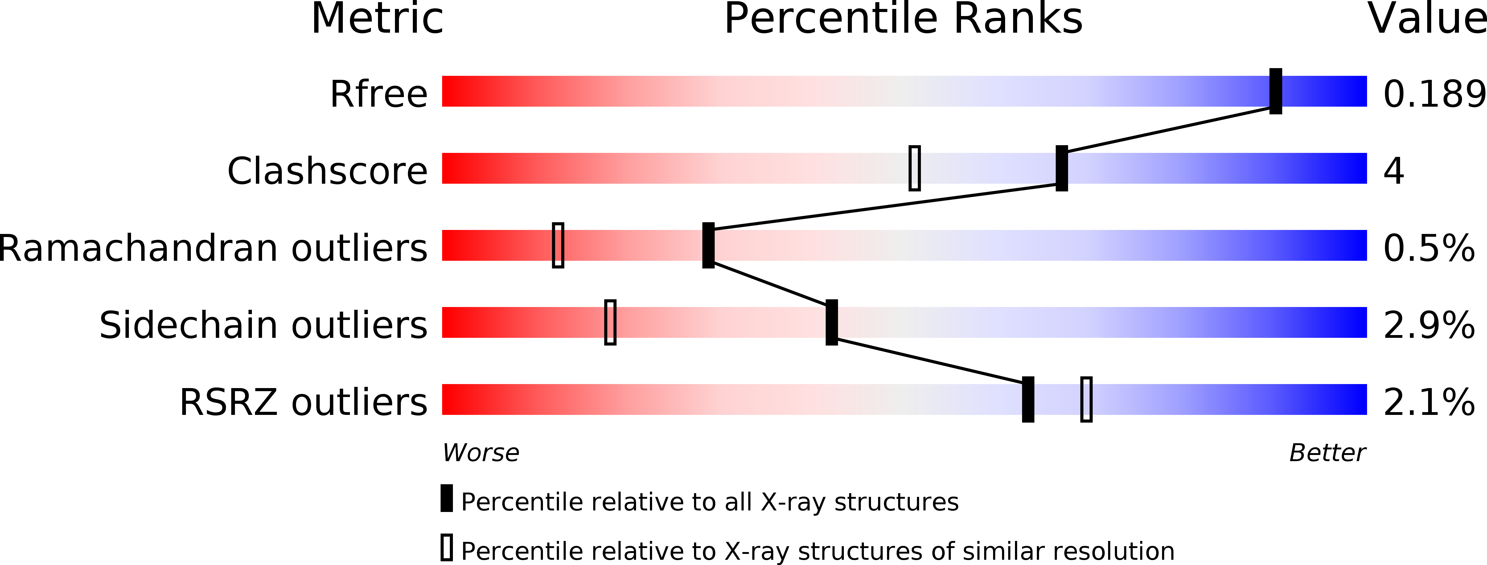
Resolution:
1.73 Å
R-Value Free:
0.18
R-Value Work:
0.15
R-Value Observed:
0.15
Space Group:
P 1 21 1
Macromolecular Entities
Polymer Type:polypeptide(L)
Description:AztD protein
Chain IDs:A (auth: B), B (auth: A)
Chain Length:421
Number of Molecules:2
Biological Source:Citrobacter koseri (strain ATCC BAA-895 / CDC 4225-83 / SGSC4696)
Primary Citation
Crystal structures of AztD provide mechanistic insights into direct zinc transfer between proteins.
Commun Biol 2 308 308 (2019)
PMID: 31428696 DOI: 10.1038/s42003-019-0542-z

Abstact

Zinc acquisition from limited environments is critical for bacterial survival and pathogenesis. AztD has been identified as a periplasmic or cell surface zinc-binding protein in numerous bacterial species. In Paracoccus denitrificans, AztD can transfer zinc directly to AztC, the solute binding protein for a zinc-specific ATP-binding cassette transporter system, suggesting a role in zinc acquisition and homeostasis. Here, we present the first cry stal structures of AztD from P. denitrificans and tbe human pathogen Citrobacter koseri, revealing a beta-propeller fold and two high-affinity zinc-binding sites that are highly conserved among AztD homologs. These structures combined with transfer assays using WT and mutant proteins provide rare insight into the mechanism of direct zinc transfer from one protein to another. Given the importance of zinc import to bacterial pathogenesis, these insights may prove valuable to the development of zinc transfer inhibitors as antibiotics.

Legend

Protein

Chemical

Disease

Primary Citation of related structures