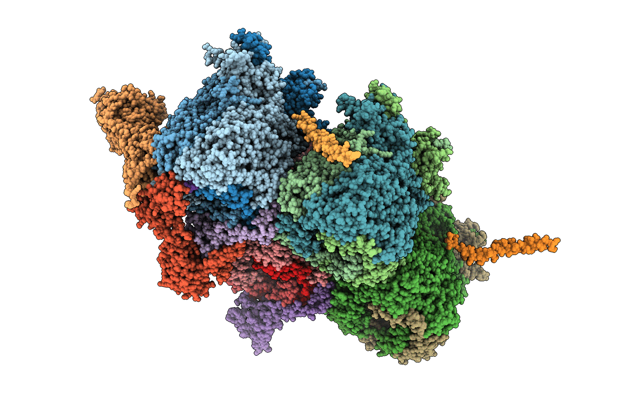
6CGV image
Entry Detail
PDB ID:
6CGV
Keywords:
Title:
Revised crystal structure of human adenovirus
Biological Source:
Host Organism:
PDB Version:
Deposition Date:
2018-02-21
Release Date:
2018-04-25
Method Details:
Experimental Method:
Resolution:
3.80 Å
R-Value Free:
0.42
R-Value Work:
0.42
Space Group:
P 1
Macromolecular Entities
Polymer Type:polypeptide(L)
Description:Hexon protein
Chain IDs:A, B, C, D, E, F, G, H, I, J, K, L
Chain Length:949
Number of Molecules:12
Biological Source:Human adenovirus C serotype 5
Polymer Type:polypeptide(L)
Description:Pre-hexon-linking protein IIIa
Chain IDs:N (auth: M)
Chain Length:585
Number of Molecules:1
Biological Source:Human adenovirus C serotype 5
Polymer Type:polypeptide(L)
Description:Penton protein
Chain IDs:M (auth: N)
Chain Length:571
Number of Molecules:1
Biological Source:Human adenovirus C serotype 5
Polymer Type:polypeptide(L)
Description:Hexon-interlacing protein
Chain IDs:O (auth: P), P (auth: Q), Q (auth: R), R (auth: S)
Chain Length:140
Number of Molecules:4
Biological Source:Human adenovirus C serotype 5
Polymer Type:polypeptide(L)
Description:Pre-hexon-linking protein VIII
Chain IDs:S (auth: U), T (auth: V)
Chain Length:227
Number of Molecules:2
Biological Source:Human adenovirus C serotype 5
Polymer Type:polypeptide(L)
Description:Pre-protein VI
Chain IDs:U (auth: W)
Chain Length:24
Number of Molecules:1
Biological Source:Human adenovirus C serotype 5
Ligand Molecules
Primary Citation
Revised Crystal Structure of Human Adenovirus Reveals the Limits on Protein IX Quasi-Equivalence and on Analyzing Large Macromolecular Complexes.
J. Mol. Biol. 430 4132 4141 (2018)
PMID: 30121295 DOI: 10.1016/j.jmb.2018.08.011

Abstact

We report the revised crystal structure of a pseudo-typed human adenovirus at 3.8-Å resolution that is consistent with the atomic models of minor proteins determined by cryo-electron microscopy. The diffraction data from multiple crystals were rescaled and merged to increase the data completeness. The densities for the minor proteins were initially identified in the phase-refined omit maps that were further improved by the phases from docked poly-alanine models to build atomic structures. While the trimeric fiber molecules are disordered due to flexibility and imposition of 5-fold symmetry, the remaining major capsid proteins hexon and penton base are clearly ordered, with the exception of hypervariable region 1 of hexons, the RGD containing loop, and the N-termini of the penton base. The exterior minor protein IX together with the interior minor proteins IIIa and VIII stabilizes the adenovirus virion. A segment of N-terminal pro-peptide of VI is found in the interior cavities of peripentonal hexons, and the rest of VI is disordered. While the triskelion substructures formed by the N-termini of IX conform to excellent quasi 3-fold symmetry, the tetrameric coiled-coils formed by the C-termini and organized in parallel and anti-parallel arrangement do not exhibit any quasi-symmetry. This observation also conveys the pitfalls of using the quasi-equivalence as validation criteria for the structural analysis of extended (non-modular) capsid proteins such as IX. Together, these results remedy certain discrepancies in the previous X-ray model in agreement with the cryo-electron microscopy models.

Legend

Protein

Chemical

Disease

Primary Citation of related structures